. 2004 Dec 15;384(Pt 3):577-84.
doi: 10.1042/BJ20041082.

Probing the active site of YjeE: a vital Escherichia coli protein of unknown function

Affiliations

Probing the active site of YjeE: a vital Escherichia coli protein of unknown function

Abdellah Allali-Hassani et al. Biochem J. .

Abstract

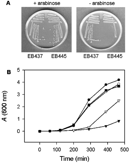
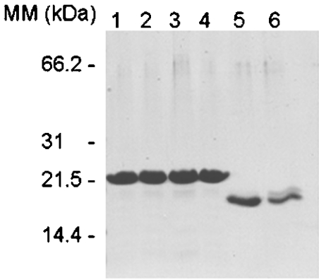
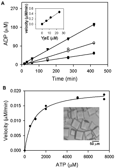
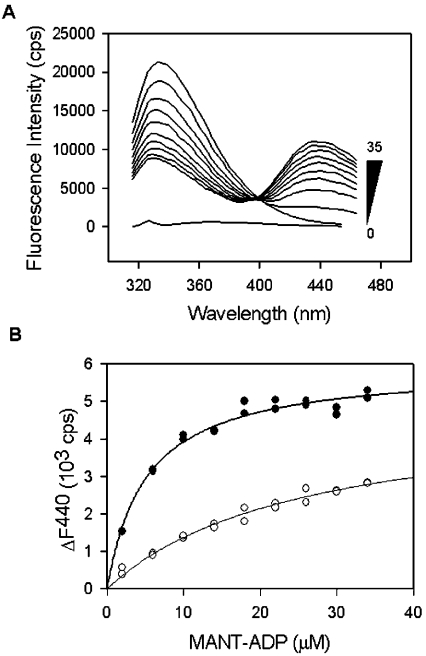
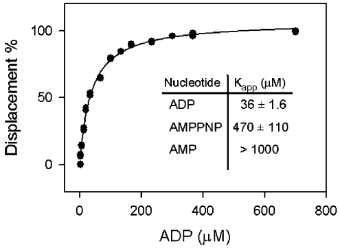
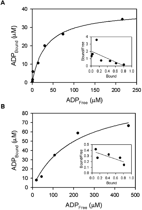
In the study described here, we have taken steps to characterize the YjeE protein, an Escherichia coli protein of unknown function that is essential for bacterial viability. YjeE represents a protein family whose members are broadly conserved in bacteria, absent from eukaryotes and contain both Walker A and B motifs, characteristic of P-loop ATPases. We have revisited the dispensability of the yjeE gene in E. coli and describe efforts to probe the function of the YjeE protein with in vitro biochemistry. We have looked critically for ATPase activity in the recombinant E. coli protein and have made vigilant use of site-directed variants in the Walker A [K41A (Lys41-->Ala) and T42A] and putative Walker B (D80Q) motifs. We noted that any hydrolysis of ATP by the wild-type E. coli protein might be attributed to background ATPase, since it was not appreciably different from that of the variants. To overcome potential contaminants, we turned to crystalline pure YjeE protein from Haemophilus influenzae that was found to hydrolyse ATP at a slow rate (kcat=1 h(-1)). We have also shown high-affinity binding to YjeE by ADP using equilibrium dialysis (K(d)=32 microM) and by fluorescence resonance energy transfer from a conserved tryptophan in YjeE to a fluorescent derivative of ADP, 2'-/3'-O-(N-methylanthraniloyl)adenosine 5'-O-diphosphate (K(d)=8 microM). Walker motif variants were notably impaired for ADP binding and T42A and D80Q mutations in yjeE were incapable of complementing the yjeE deletion strain.


Figures

Figure 1
Figure 1. Conditional complementation of the yjeE deletion strain
(A) Arabinose dependence of the yjeE deletion strain EB445. The yjeE deletion strain (EB445) and the diploid strain (EB437) were plated in the presence and absence of arabinose and grown overnight at 37 °C. (B) Growth of yjeE-depleted cells in liquid media. Cells (EB445) were grown overnight on LB/KAN/arabinose plates and used to inoculate LB/KAN media with 0.2% (■), 0.02% (▽) and no arabinose (▼). The diploid strain (EB437) was similarly grown in the presence of 0.2% arabinose (●) and no arabinose (○). Growth was followed at 37 °C for 8 h.
Figure 2
Figure 2. SDS/PAGE (15% gel) showing purified YjeE and its variants
Lane 1, wild-type E. coli YjeE; lane 2, E. coli K41A variant; lane 3, E. coli T42A variant; lane 4, E. coli D80Q variant; lanes 5 and 6, purified recombinant H. influenzae YjeE before and after crystallization respectively.
Figure 3
Figure 3. ATPase activity of crystalline pure H. influenzae YjeE
Crystals of YjeE (B, inset) were prepared and dissolved as described (in the Experimental section) and ATPase activity was determined. (A) YjeE at concentrations of 6 μM (●), 12 μM (○) and 24 μM (▼) was incubated in the presence of 6 mM ATP and 10 mM MgCl2 (reaction volume, 100 μl) and 10 μl fractions were sampled at the times indicated and quenched by the addition of urea to 6 M. The amount of ADP produced was determined by HPLC as described in the Experimental section. The inset shows the initial velocity of the reaction plotted as a function of enzyme concentration; the slope represents the turnover of the enzyme under these conditions (1.2 h−1). (B) Dependence of the reaction velocity on ATP concentration. Reactions (20 μl) containing ATP (0.2–7.5 mM), YjeE (12 μM) and 10 mM MgCl2 were quenched after 7 h and analysed by HPLC to determine the rates. The data were fitted to the Michaelis–Menten equation v=kcat[E][S]/(Km+[S]) using Sigma Plot 2000 (SPSS Science, Chicago, IL, U.S.A.) and Km and kcat values were calculated to be 800 μM and 1.2 h−1 respectively.
Figure 4
Figure 4. Determination of Kd for wild-type YjeE and the K41A variant
(A) Emission spectra collected on incubation of recombinant E. coli YjeE protein (2 μM) with increasing amounts of MANT-ADP (0–35 μM) as indicated (excitation was at 295 nm). The spectrum shown at the bottom corresponds to that of buffer alone. (B) Change of fluorescence emission intensity at 440 nm for wild-type YjeE (●) and the K41A variant (○) as a function of MANT-ADP concentration. The spectra of the protein alone and MANT-ADP alone at each concentration were subtracted from the spectra of MANT-ADP in the presence of protein, and the change of fluorescence at 440 nm (ΔF440) was plotted as a function of the total MANT-ADP concentration. Fluorescence is expressed in terms of counts/s (cps). The data were fitted to a hyperbolic function to determine Kd values of 8 and 65 μM for wild-type YjeE and the K41A variant respectively.
Figure 5
Figure 5. Displacement of MANT-ADP bound to YjeE by various nucleotides
The graph depicts the displacement of the MANT-ADP by unmodified ADP as followed by a decrease in the FRET signal associated with the binding of the MANT-ADP (10 μM) to YjeE (6 μM). The data were fitted to a hyperbolic function to determine the Kapp. The inset summarizes the Kapp measured for ADP, AMPPNP and AMP. No displacement was detected when GMP, GDP, GTP, CMP, CDP, CTP, UMP, UDP or UTP was used as a competitor.
Figure 6
Figure 6. Determination of Kd for ADP bound to wild-type YjeE (A) and K41A variant (B) using equilibrium dialysis
The data are presented in hyperbolic form, where [ADP]Bound is plotted versus [ADP]Free. Equilibration was attained as described in the Experimental section under the following conditions: 20 mM Tris, 2 mM DTT, 10 mM MgCl2 (pH 7.5) and 33 μM wild-type YjeE and 100 μM K41A variant respectively. The insets show Scatchard plots where the [ADP]Bound/[ADPFree] ratio is plotted as a function of [ADP]Bound. The stoichiometry of binding shows that about one molecule of ADP is bound per molecule of protein. The Kd values for ADP bound to wild-type YjeE and K41A variant were calculated to be 32 and 211 μM respectively.
Figure 7
Figure 7. In vivo complementation of yjeE deletion strain with wild-type and variants of YjeE
(A) Strains were grown on LB/CM/arabinose plates (left panel) and LB/CM plates (right panel) at 37 °C overnight. Clockwise from top: strain EB445 (yjeE deletion strain), EB445 with pDEST17-WTYjeE, EB445 with pDEST17-K41AYjeE, EB445 with pDEST17-T42AYjeE and EB445 with pDEST17-D80QYjeE. (B) Western blotting using a YjeE polyclonal antibody to confirm expression of YjeE from the pDEST17 plasmids. Cell lysates (approx. 100 μg) were subjected to SDS/PAGE (15% gel) and electroblotted on to a nitrocellulose membrane. The blot was treated with YjeE polyclonal antibody and donkey anti-rabbit antibody conjugated to horseradish peroxidase. The signal was developed using the Western Exposure Chemiluminescent Detection System and imaged on a chemiluminescence film with a 2 min exposure. Lane 1, wild-type E. coli MG1655; lane 2, EB437 (diploid strain); lane 3, EB437/pDEST17-WTYjeE; lane 4, EB437/pDEST17-K41AYjeE; lane 5, EB437/pDEST17-T42AYjeE; lane 6, EB437/pDEST17-D80QYjeE. Evident from the blot are both His-tagged (expressed from pDEST17) and untagged YjeE migrating with apparent molecular masses of 19.2 and 18.1 kDa respectively.

References

    1. Fleischmann R. D., Adams M. D., White O., Clayton R. A., Kirkness E. F., Kerlavage A. R., Bult C. J., Tomb J. F., Dougherty B. A., Merrick J. M. Whole-genome random sequencing and assembly of Haemophilus influenzae Rd. Science. 1995;269:496–512. - PubMed
    1. Tatusov R. L., Galperin M. Y., Natale D. A., Koonin E. V. The COG database: a tool for genome-scale analysis of protein functions and evolution. Nucleic Acids Res. 2000;28:33–36. - PMC - PubMed
    1. Venter J. C., Adams M. D., Myers E. W., Li P. W., Mural R. J., Sutton G. G., Smith H. O., Yandell M., Evans C. A., Holt R. A., et al. The sequence of the human genome. Science. 2001;291:1304–1351. - PubMed
    1. Lander E. S., Linton L. M., Birren B., Nusbaum C., Zody M. C., Baldwin J., Devon K., Dewar K., Doyle M., FitzHugh W., et al. Initial sequencing and analysis of the human genome. Nature (London) 2001;409:860–921. - PubMed
    1. Badarinarayana V., Estep P. W., 3rd, Shendure J., Edwards J., Tavazoie S., Lam F., Church G. M. Selection analyses of insertional mutants using subgenic-resolution arrays. Nat. Biotechnol. 2001;19:1060–1065. - PubMed

Publication types

MeSH terms

LinkOut - more resources