Comparative Study
. 2004 Nov;87(5):3148-61.
doi: 10.1529/biophysj.104.046664. Epub 2004 Aug 23.

The external TEA binding site and C-type inactivation in voltage-gated potassium channels

Affiliations
Comparative Study

The external TEA binding site and C-type inactivation in voltage-gated potassium channels

Payam Andalib et al. Biophys J. 2004 Nov.

Abstract

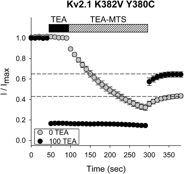
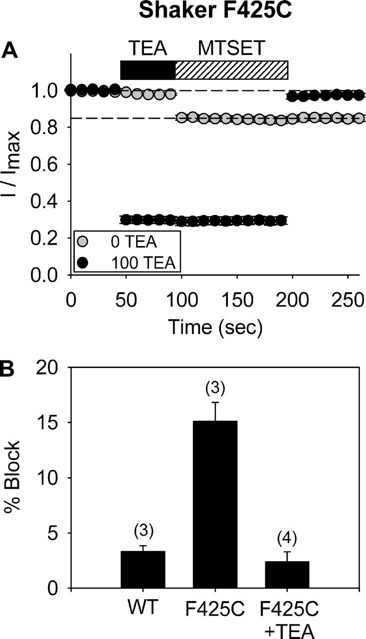
The location of the tetraethylammonium (TEA) binding site in the outer vestibule of K+ channels, and the mechanism by which external TEA slows C-type inactivation, have been considered well-understood. The prevailing model has been that TEA is coordinated by four amino acid side chains at the position equivalent to Shaker T449, and that TEA prevents a constriction that underlies inactivation via a foot-in-the-door mechanism at this same position. However, a growing body of evidence has suggested that this picture may not be entirely correct. In this study, we reexamined these two issues, using both the Kv2.1 and Shaker potassium channels. In contrast to results previously obtained with Shaker, substitution of the tyrosine at Kv2.1 position 380 (equivalent to Shaker 449) with a threonine or cysteine had a relatively minor effect on TEA potency. In both Kv2.1 and Shaker, modification of cysteines at position 380/449 by 2-(trimethylammonium)ethyl methanethiosulfonate (MTSET) proceeded at identical rates in the absence and presence of TEA. Additional experiments in Shaker demonstrated that TEA bound well to C-type inactivated channels, but did not interfere with MTSET modification of C449 in inactivated channels. Together, these findings rule out the possibility that TEA binding involves an intimate interaction with the four side chains at the position equivalent to Shaker 449. Moreover, these results argue against the model whereby TEA slows inactivation via a foot-in-the-door mechanism at position 449, and also argue against the hypothesis that the position 449 side chains move toward the center of the conduction pathway during inactivation. Occupancy by TEA completely prevented MTSET modification of a cysteine in the outer-vestibule turret (Kv2.1 position 356/Shaker position 425), which has been shown to interfere with both TEA binding and the interaction of K+ with an external binding site. Together, these data suggest that TEA is stabilized in a more external position in the outer vestibule, and does not bind via direct coordination with any specific outer-vestibule residues.


Figures

FIGURE 1
FIGURE 1
Block of Kv2.1 K382V by TEA after mutation of Y380. Concentration-dependent block by external TEA is shown for three channels: Kv2.1 K382V, which has the native tyrosine at position 380 and serves as the control channel (circles), and Kv2.1 K382V channels, which have the position 380 residue mutated to a threonine (Y380T; triangles) or a cysteine (Y380C; squares). Solid lines are best fits to the curves from Eq. 1, which gave calculated EC50 values of 0.95 mM for Y380, 5.0 mM for T380, and 13.4 mM for C380. All data points represent the mean ± SE of three cells. Recordings were made with 100 mM internal and 5 mM external K+ (NMG made up the balance of the monovalent cation).
FIGURE 2
FIGURE 2
External TEA did not affect the rate of MTSET modification of C380 in Kv2.1. (A) Reversible block of Kv2.1 K382V Y380C by 100 mM external TEA. (B) After a 50-s application of 100 mM TEA alone (marked by black bar), 100 mM TEA plus 2 mM MTSET was applied for 200 s (marked by hatched bar) to a channel without the cysteine at position 380. Data points in A and B represent the mean ± SE of four cells. (C)Four different conditions are shown. The shaded symbols illustrate the time course of modification of C380 by 2 mM MTSET with a 40-s application (triangles) and 200-s application (circles). MTSET was removed after 40 s (OFF 40) or after 200 s (OFF 200), and irreversible block measured once current relaxed to a new steady-state level. The black symbols illustrate the time course of modification by MTSET, applied for these same two durations, in the presence of 100 mM TEA (TEA was applied for 40 s before, and throughout, exposure to MTSET). At both the 40- and 200-s time points, TEA and MTSET were removed simultaneously. Data points represent the mean ± SE of 3–6 cells.
FIGURE 3
FIGURE 3
External TEA interfered with modification by TEA-MTS. The experimental protocol was the same as for Fig. 2: TEA-MTS was applied by itself (shaded circles; n = 13) or with prior and simultaneous application of 100 mM TEA (black circles; n = 5).
FIGURE 4
FIGURE 4
External TEA potency after modification of C380 by MTSET or TEA-MTS. (A) Black circles illustrate TEA concentration dependence without MTSET treatment (data are identical to those shown in Fig. 1). Shaded circles illustrate TEA concentration dependence after a 3-min preincubation with 2 mM MTSET. The IC50s for block by TEA, calculated from the best fits to Eq. 1, were 13.4 mM without MTSET and 88.7 mM after MTSET preincubation. Essentially identical results were obtained when an alternative protocol, used for panel B experiments, was used (data not shown). (B) TEA concentration dependence was determined in the absence of TEA-MTS treatment (black circles). Then, on the same cells, 2 mM TEA-MTS was applied for 3 min, and the TEA concentration dependence was determined again. Experiments in panel B were done with 0 mM external K+. All data points in panels A and B represent the mean ± SE of 3–4 cells.
FIGURE 5
FIGURE 5
Influence of external TEA on modification of C380 by MTSPT and MTSBT. (A) Illustration of structures of four MTS reagents. Length of R group is shown after the name of the compound. (B and C) Modification of C380 by MTSPT (B) and MTSBT (C) in the absence and presence of 100 mM external TEA. Experimental protocol are as in Fig. 2. Data points represent mean ± SE of three cells.
FIGURE 6
FIGURE 6
External TEA protects C356 from modification by MTSET. (A) Irreversible block of current after exposure of Kv2.1 K382V K356C to 2 mM MTSET (n = 4). (B) Prior and simultaneous application of 100 mM TEA completely prevented irreversible block of channel by MTSET (n = 4). (C) I-V relationships constructed under three different conditions, with 125 mM internal and external Na+ plus 1 mM external K+. In the absence of MTSET treatment, currents reversed at 11.4 mV. After a 3-min preincubation with 2 mM MTSET, the reversal potential shifted to 3.4 mV, which reflected modification of C356 (see Consiglio et al., 2003). Simultaneous preincubation with 100 mM TEA completely prevented the reversal potential shift associated with MTSET modification of C356 (Erev = 12.3 mV). Data are illustrated from three individual cells, one in each condition. (D) Composite data from several cells, examined as in panel C (value in parentheses represents number of cells tested). The reversal potential values from cells not exposed to MTSET and from cells pretreated simultaneously with TEA and MTSET were statistically identical (11.7 ± 0.4 mV and 12.0 ± 0.6 mV, respectively).
FIGURE 7
FIGURE 7
TMA has no functional effect on the channel. 100 mM TMA was applied in the absence or presence of 3 mM TEA. Data represent the mean ± SE of three cells in each condition.
FIGURE 8
FIGURE 8
Modification of C449 in Shaker by MTSET. (A) Outward currents after 50-ms depolarizations to 0 mV in control and after application of 300 μM MTSET for various durations. (B) Time course of current block by MTSET in the absence (shaded circles; n = 4) and presence (black circles; n = 3) of 100 mM external TEA. Protocol was the same as in Fig. 2, except that MTSET was applied for 150 s. (C) Three superimposed currents are shown, evoked by depolarization to 0 mV for 14 s. In the first trace, no MTSET was applied. In the second trace, 300 μM MTSET was applied 8 s after the start of the depolarization (arrow) and left on the cell until the end of the stimulus, for a total application duration of 6 s. The third (smaller) current was recorded 10 s after MTSET removal. (D) Protocol was identical to that in panel C, except that 100 mM TEA was applied 1 s before and for the duration of MTSET application. (E) Plot of normalized peak current, measured from five consecutive traces. MTSET was applied for 6 s during the third trace, in the absence (circles; n = 3) and presence (squares; n = 3) of TEA. (F) Plot of the change in the inactivation time constant by MTSET in the absence and presence of TEA, obtained from the same data as in panel E. The effect of MTSET was statistically identical under the two conditions (2.4 ± 0.3 s and 2.4 ± 0.1 s, respectively; n = 3 for each).
FIGURE 9
FIGURE 9
Modification of C449 in Shaker by TEA-MTS. Protocols and plot construction in each panel are identical to those in Fig. 8, except that C449 was modified by TEA-MTS. Current block (panels B and E) and change in inactivation time constant (panel F) were significantly reduced by the simultaneous presence of TEA during TEA-MTS exposure.
FIGURE 10
FIGURE 10
TEA interferes with MTSET modification of C425 in Shaker. (A) MTSET (300 μM) was applied in the absence (shaded circles) and presence (black circles) of 100 mM TEA. Data points represent mean ± SE of 3–4 cells. (B) Composite data for three conditions, including the wild-type channel (WT), which lacked the cysteine in the outer vestibule. All cells were preincubated for 3 min with 10 mM DTT.
FIGURE 11
FIGURE 11
Models of TEA interaction with the outer vestibule. All models were created with Swiss-PdbViewer and edited with RasTop, using the KcsA backbone, threaded with Kv2.1 residues and minimized. (A and B) Top-down view of the channel with cysteine residues (panel A, blue) or tyrosine residues (panel B, red) illustrated at position 380. Arrows in panel A point to the reactive sulfur group on the cysteine side chain. TEA is shown in the conduction pathway (yellow). Note that the cysteine reactive side chains are pointed down and away from the central axis of the conduction pathway (A), whereas the tyrosine side-chain aromatics are pointed up and toward the center of the conduction pathway (B). (C) Previous model of TEA binding to the channel. View of channel is from the side, with two of four subunits shown. For illustration purposes, the channel is depicted with cysteine residues at position 356 in the turret and at position 380. The selectivity filter GYG residues are also illustrated as stick representations. Arrows point to reactive cysteine side chain at position 356. (D) Proposed new model, wherein TEA is stabilized more externally in the outer vestibule and placed so as not to act as a foot in the door in a constricting selectivity filter. (E) Same as panel D, except that tyrosine residues (red) are illustrated at position 380. The TEA molecule in these figures was reproduced from The Journal of General Physiology, 2001, 118:207–217, by copyright permission of The Rockefeller University Press.

References

    1. Andalib, P., M. J. Wood, and S. J. Korn. 2002. Control of outer vestibule dynamics and current magnitude in the Kv2.1 potassium channel. J. Gen. Physiol. 120:739–755. - PMC - PubMed
    1. Armstrong, C. M. 1969. Inactivation of the potassium conductance and related phenomena caused by quaternary ammonium ion injection in squid axons. J. Gen. Physiol. 54:553–575. - PMC - PubMed
    1. Baukrowitz, T., and G. Yellen. 1996. Use-dependent blockers and exit rate of the last ion from the multi-ion pore of a K+ channel. Science. 271:653–656. - PubMed
    1. Blaustein, R. O., P. A. Cole, C. Williams, and C. Miller. 2000. Tethered blockers as molecular ‘tape measures’ for a voltage-gated K+ channel. Nat. Struct. Biol. 7:309–311. - PubMed
    1. Bretschneider, F., A. Wrisch, F. Lehmann-Horn, and S. Grissmer. 1999. External tetraethylammonium as a molecular caliper for sensing the shape of the outer vestibule of potassium channels. Biophys. J. 76:2351–2360. - PMC - PubMed

Publication types

MeSH terms

Substances

LinkOut - more resources