

Identification of an alternative ligand-binding pocket in the nuclear vitamin D receptor and its functional importance in 1alpha,25(OH)2-vitamin D3 signaling
- PMID: 15326291
- PMCID: PMC516488
- DOI: 10.1073/pnas.0403606101
Identification of an alternative ligand-binding pocket in the nuclear vitamin D receptor and its functional importance in 1alpha,25(OH)2-vitamin D3 signaling
Abstract
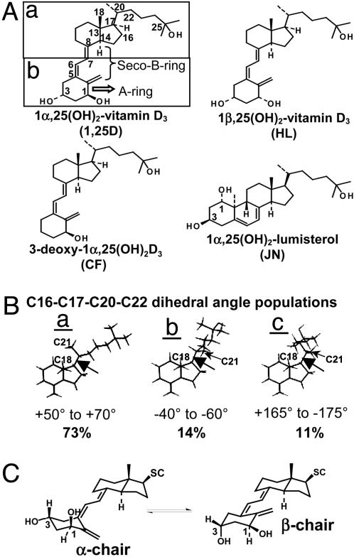
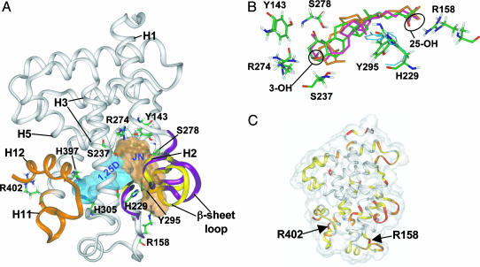
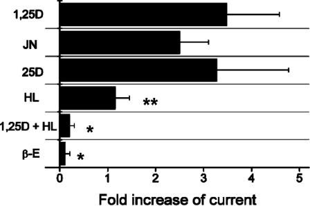
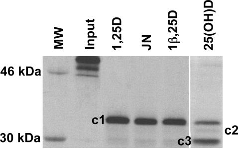
Structural and molecular studies have shown that the vitamin D receptor (VDR) mediates 1alpha,25(OH)2-vitamin D3 gene transactivation. Recent evidence indicates that both VDR and the estrogen receptor are localized to plasma membrane caveolae and are required for initiation of nongenomic (NG) responses. Computer docking of the NG-specific 1alpha,25(OH)2-lumisterol to the VDR resulted in identification of an alternative ligand-binding pocket that partially overlaps the genomic pocket described in the experimentally determined x-ray structure. Data obtained from docking five different vitamin D sterols in the genomic and alternative pockets were used to generate a receptor conformational ensemble model, providing an explanation for how VDR and possibly the estrogen receptor can have genomic and NG functionality. The VDR model is compatible with the following: (i) NG chloride channel agonism and antagonism; (ii) variable ligand-stabilized trypsin digest banding patterns; and (iii) differential transcriptional activity, employing different VDR point mutants and 1alpha,25(OH)2-vitamin D3 analogs.
Copyright 2004 The National Academy of Sciencs of the USA
Figures
Similar articles
-
Applications of the Vitamin D sterol-Vitamin D receptor (VDR) conformational ensemble model.Steroids. 2005 May-Jun;70(5-7):464-71. doi: 10.1016/j.steroids.2005.03.003. Epub 2005 Apr 8. Steroids. 2005. PMID: 15862832
-
Vitamin D receptor (VDR)-mediated actions of 1α,25(OH)₂vitamin D₃: genomic and non-genomic mechanisms.Best Pract Res Clin Endocrinol Metab. 2011 Aug;25(4):543-59. doi: 10.1016/j.beem.2011.05.010. Best Pract Res Clin Endocrinol Metab. 2011. PMID: 21872797 Review.
-
A perspective on how the Vitamin D sterol/Vitamin D receptor (VDR) conformational ensemble model can potentially be used to understand the structure-function results of A-ring modified Vitamin D sterols.J Steroid Biochem Mol Biol. 2005 Oct;97(1-2):69-82. doi: 10.1016/j.jsbmb.2005.06.025. Epub 2005 Aug 1. J Steroid Biochem Mol Biol. 2005. PMID: 16055325
-
25-Dehydro-1alpha-hydroxyvitamin D3-26,23S-lactone antagonizes the nuclear vitamin D receptor by mediating a unique noncovalent conformational change.Mol Endocrinol. 2000 Nov;14(11):1788-96. doi: 10.1210/mend.14.11.0552. Mol Endocrinol. 2000. PMID: 11075812
-
Minireview: vitamin D receptor: new assignments for an already busy receptor.Endocrinology. 2006 Dec;147(12):5542-8. doi: 10.1210/en.2006-0946. Epub 2006 Aug 31. Endocrinology. 2006. PMID: 16946007 Review.
Cited by
-
The Role of Classical and Novel Forms of Vitamin D in the Pathogenesis and Progression of Nonmelanoma Skin Cancers.Adv Exp Med Biol. 2020;1268:257-283. doi: 10.1007/978-3-030-46227-7_13. Adv Exp Med Biol. 2020. PMID: 32918223 Free PMC article. Review.
-
Evidence for Involvement of Nonclassical Pathways in the Protection From UV-Induced DNA Damage by Vitamin D-Related Compounds.JBMR Plus. 2021 Sep 29;5(12):e10555. doi: 10.1002/jbm4.10555. eCollection 2021 Dec. JBMR Plus. 2021. PMID: 34950826 Free PMC article.
-
Identification of novel immunoregulatory molecules in human thymic regulatory CD4+CD25+ T cells by phage display.PLoS One. 2011;6(8):e21702. doi: 10.1371/journal.pone.0021702. Epub 2011 Aug 1. PLoS One. 2011. PMID: 21829599 Free PMC article.
-
Structural considerations of vitamin D signaling.Front Physiol. 2014 Jun 6;5:191. doi: 10.3389/fphys.2014.00191. eCollection 2014. Front Physiol. 2014. PMID: 24936188 Free PMC article. Review.
-
The role of vitamin D in the endocrinology controlling calcium homeostasis.Mol Cell Endocrinol. 2017 Sep 15;453:36-45. doi: 10.1016/j.mce.2017.04.008. Epub 2017 Apr 9. Mol Cell Endocrinol. 2017. PMID: 28400273 Free PMC article. Review.
References
-
- Losel, R. & Wehling, M. (2003) Nat. Rev. Mol. Cell Biol. 4, 46–56. - PubMed
-
- Norman, A. W., Mizwicki, M. T. & Norman, D. P. G. (2004) Nat. Rev. Drug Discov. 3, 27–41. - PubMed
-
- Caffrey, J. M. & Farach-Carson, M. C. (1989) J. Biol. Chem. 264, 20265–20274. - PubMed
-
- Zanello, L. P. & Norman, A. W. (1997) J. Biol. Chem. 272, 22617–22622. - PubMed
Publication types
MeSH terms
Substances
Grants and funding
LinkOut - more resources
Full Text Sources
Other Literature Sources
