. 2004 Sep;48(9):3291-7.
doi: 10.1128/AAC.48.9.3291-3297.2004.

Penetration of Candida biofilms by antifungal agents

Affiliations

Penetration of Candida biofilms by antifungal agents

Mohammed A Al-Fattani et al. Antimicrob Agents Chemother. 2004 Sep.

Abstract

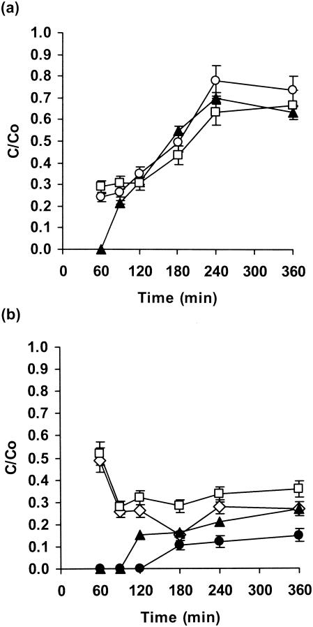
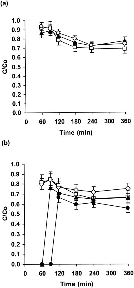
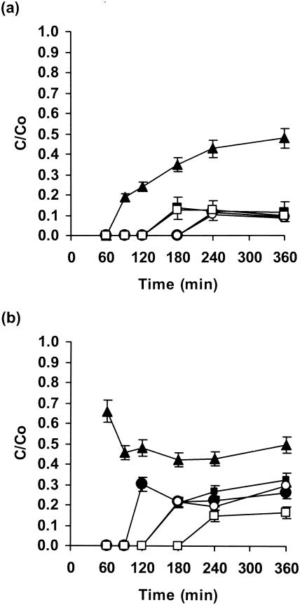
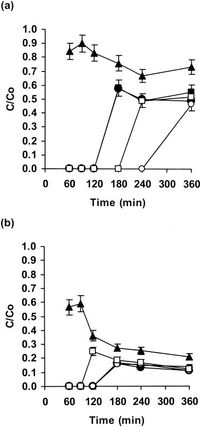
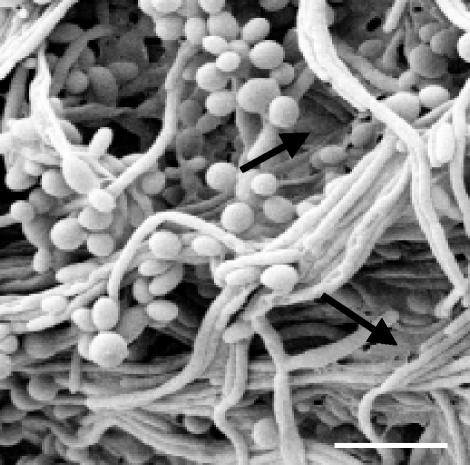
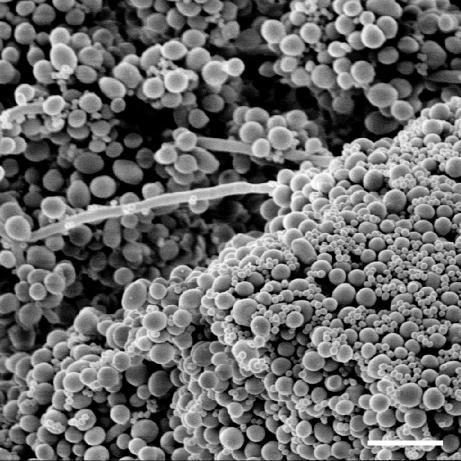
A filter disk assay was used to investigate the penetration of antifungal agents through biofilms containing single and mixed-species biofilms containing Candida. Fluconazole permeated all single-species Candida biofilms more rapidly than flucytosine. The rates of diffusion of either drug through biofilms of three strains of Candida albicans were similar. However, the rates of drug diffusion through biofilms of C. glabrata or C. krusei were faster than those through biofilms of C. parapsilosis or C. tropicalis. In all cases, after 3 to 6 h the drug concentration at the distal edge of the biofilm was very high (many times the MIC). Nevertheless, drug penetration failed to produce complete killing of biofilm cells. These results indicate that poor antifungal penetration is not a major drug resistance mechanism for Candida biofilms. The abilities of flucytosine, fluconazole, amphotericin B, and voriconazole to penetrate mixed-species biofilms containing C. albicans and Staphylococcus epidermidis (a slime-producing wild-type strain, RP62A, and a slime-negative mutant, M7) were also investigated. All four antifungal agents diffused very slowly through these mixed-species biofilms. In most cases, diffusion was slower with biofilms containing S. epidermidis RP62A, but amphotericin B penetrated biofilms containing the M7 mutant more slowly. However, the drug concentrations reaching the distal edges of the biofilms always substantially exceeded the MIC. Thus, although the presence of bacteria and bacterial matrix material undoubtedly retarded the diffusion of the antifungal agents, poor penetration does not account for the drug resistance of Candida biofilm cells, even in these mixed-species biofilms.


Figures

FIG. 1.
FIG. 1.
Penetration of flucytosine through biofilms of C. albicans GDH 2346 (closed triangles), C. albicans GDH 2023 (open squares), and C. albicans GRI 682 (open circles) (a) and through biofilms of C. krusei (open squares), C. glabrata (open diamonds), C. parapsilosis (closed triangles), and C. tropicalis (closed circles) (b). Error bars indicate the standard errors of the means. The mean C0 after 6 h was 22.7 μg of flucytosine/ml.
FIG. 2.
FIG. 2.
Penetration of fluconazole through biofilms of C. albicans strain GDH 2346 (closed triangles), C. albicans GDH 2023 (open squares), and C. albicans GRI 682 (open circles) (a) and through biofilms of C. krusei (open squares), C. glabrata (open diamonds), C. parapsilosis (closed triangles), and C. tropicalis (closed circles) (b). Error bars indicate the standard errors of the means. The mean C0 after 6 h was 26.6 μg of fluconazole/ml.
FIG. 3.
FIG. 3.
Penetration of flucytosine (a) and amphotericin B (b) through single- and mixed-species biofilms of C. albicans and S. epidermidis. Biofilms contained C. albicans GDH 2346 (closed triangles), S. epidermidis RP62A (closed circles), S. epidermidis M7 (closed squares), C. albicans GDH 2346 and S. epidermidis RP62A (open circles), and C. albicans GDH 2346 and S. epidermidis M7 (open squares). Error bars indicate the standard errors of the means. Mean C0s after 6 h were 24.2 μg of flucytosine/ml and 120.6 μg of amphotericin B/ml.
FIG. 4.
FIG. 4.
Penetration of fluconazole (a) and voriconazole (b) through single- and mixed-species biofilms of C. albicans and S. epidermidis. Biofilms contained C. albicans GDH 2346 (closed triangles), S. epidermidis RP62A (closed circles), S. epidermidis M7 (closed squares), C. albicans GDH 2346 and S. epidermidis RP62A (open circles), and C. albicans GDH 2346 and S. epidermidis M7 (open squares). Error bars indicate the standard errors of the means. Mean C0s after 6 h were 27.0 μg of fluconazole/ml and 7.9 μg of voriconazole/ml.
FIG. 5.
FIG. 5.
Scanning electron micrograph of a 48-h-old colony (membrane-supported) biofilm of C. tropicalis. Arrows indicate extracellular matrix material. Bar, 10 μm.
FIG. 6.
FIG. 6.
Scanning electron micrograph of a 48-h-old colony (membrane-supported) biofilm of C. albicans GDH 2346 and S. epidermidis M7. Bar, 10 μm.

References

    1. Adam, B., G. S. Baillie, and L. J. Douglas. 2002. Mixed species biofilms of Candida albicans and Staphylococcus epidermidis. J. Med. Microbiol. 51:344-349. - PubMed
    1. Allison, D. G., and M. J. Matthews. 1992. Effect of polysaccharide interactions on antibiotic susceptibility of Pseudomonas aeruginosa. J. Appl. Bacteriol. 73:484-488. - PubMed
    1. Anderl, J. N., M. J. Franklin, and P. S. Stewart. 2000. Role of antibiotic penetration limitation in Klebsiella pneumoniae biofilm resistance to ampicillin and ciprofloxacin. Antimicrob. Agents Chemother. 44:1818-1824. - PMC - PubMed
    1. Bachmann, S. P., K. VandeWalle, G. Ramage, T. F. Patterson, B. L. Wickes, J. R. Graybill, and J. L. Lopez-Ribot. 2002. In vitro activity of caspofungin against Candida albicans biofilms. Antimicrob. Agents Chemother. 46:3591-3596. - PMC - PubMed
    1. Baillie, G. S., and L. J. Douglas. 1998. Effect of growth rate on resistance of Candida albicans biofilms to antifungal agents. Antimicrob. Agents Chemother. 42:1900-1905. - PMC - PubMed

Publication types

MeSH terms

LinkOut - more resources