. 2005 Jan 1;105(1):233-40.
doi: 10.1182/blood-2004-03-1075. Epub 2004 Aug 24.

NKG2D receptor-mediated NK cell function is regulated by inhibitory Ly49 receptors

Affiliations

NKG2D receptor-mediated NK cell function is regulated by inhibitory Ly49 receptors

Jeyarani Regunathan et al. Blood. .

Abstract

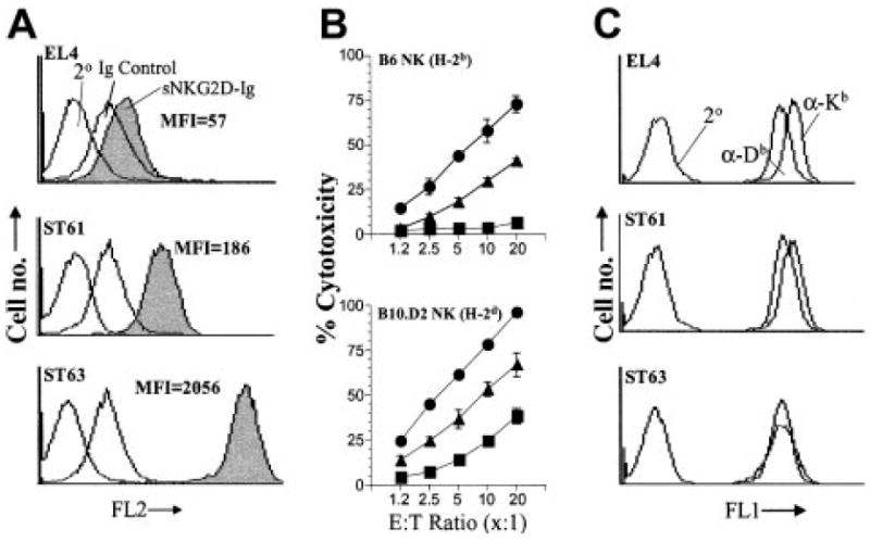
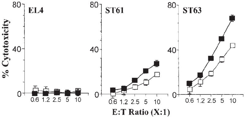
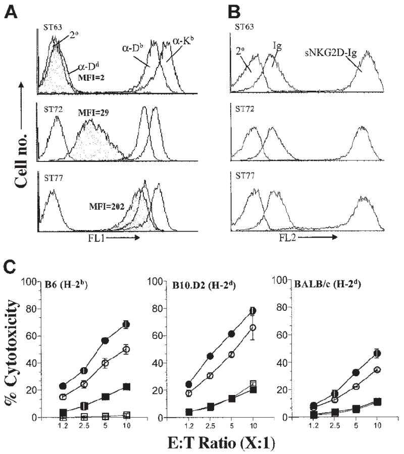
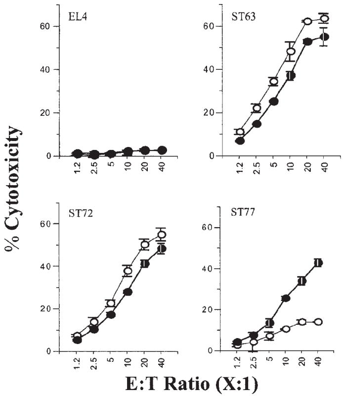
Interaction of the activating ligand H60 with NKG2D receptor constitutes a major stimulatory pathway for natural killer (NK) cells. The influence of inhibitory Ly49 receptors on NKG2D-mediated activation is not clearly understood. Here we show that the magnitude of NKG2D-mediated cytotoxicity is directly proportional to both the levels of H60 and the nature of major histocompatibility complex (MHC) class I molecules expressed on the target cells. The expression levels of H60 on the target cells determined the extent to which the inhibition by Ly49C/I receptors can be overridden. In contrast, even a higher expression of H60 molecule on the target cells failed to overcome the inhibition mediated by Ly49A/G receptors. Also, the level of interferon-gamma (IFN-gamma) and granulocyte-macrophage colony-stimulating factor (GM-CSF) generated by NK cells through anti-NKG2D monoclonal antibody (mAb)-mediated activation is significantly reduced by the presence of immobilized anti-Ly49A/G mAbs. Thus, NKG2D-mediated cytotoxicity and cytokine secretion results from the fine balance between activating and inhibitory receptors, thereby defining the NK cell-mediated immune responses.


Figures

Figure 1
Figure 1. Expression level of H60 is directly proportional to the level of NKG2D-mediated activation
(A) Expression levels of H60 on target cells were quantified by flow cytometry following staining with sNKG2D-Ig fusion protein. Soluble immunoglobulin Fc (Ig) or the secondary antibody alone (2°) was used to determine background staining levels. (B) Cytotoxicity of B6- and B10.D2-derived NK cells against EL4, ST61, and ST63. Increasing numbers of NK cells were titrated against a constant number of 51Cr-labeled target cells (5000 per well), and percent cytotoxicity was determined in a conventional 4-hour cytotoxicity assay. Data presented as the mean percent cytotoxicity ± standard deviation (SD) were determined from triplicate wells and are representative of 6 independent experiments. (C) Expression levels of MHC class I molecules in EL4, ST61, and ST63 were tested using anti–H2-Kb (α-Kb) and anti–H2-Db (α-Db) mAbs.
Figure 2
Figure 2. Differential expression levels of H60 influence the Ly49C/I receptor–mediated inhibition
(A) The effect of inhibitory Ly49 receptors on cytotoxicity was tested using Ly49C/I-specific SW5E6 or (B) Ly49I-specific YLI-90 mAbs at titrated concentrations (ST63 ●, ST61 ▲, and EL4 ■; respective open symbols denote no antibody) and (C-D) with different E / T ratios using constant amounts of mAbs (2 μg per well) (histograms). Note different Y-axis scales for ST61 and ST63 data. Results presented as the mean percent cytotoxicity ± SD and are representative of 3 independent experiments.
Figure 3
Figure 3. Expression levels of H60 differentially activate purified Ly49C+/ I+ compared with Ly49C/I NK subsets
The effect of Ly49C/I receptors on NK cell–mediated cytotoxicity was tested as described in the legend to Figure 1B using sorted subsets of Ly49C+/I+ and Ly49C/I NK cells and are representative of 3 independent experiments. Data presented are the mean values of percent cytotoxicity against indicated target cells with Ly49C+/I+ (□) or Ly49C/I (■).
Figure 4
Figure 4. Coexpression of H2-Dd on H60-positive target cells significantly inhibits NK cell cytotoxicity
(A) ST63 (H60hi) cells were transfected with H2-Dd–encoding plasmids to obtain sublines expressing low (ST72) and high (ST77) levels of H2-Dd. Expression levels of MHC class I were quantified by flow cytometric analysis following staining with anti-Dd (α-Dd), anti-Kb (α-Kb), and anti-Db (α-Db) mAbs. Secondary antibody alone (2°) was used to determine background staining levels. (B) Comparable levels of H60 on ST63, ST72, and ST77 were verified by flow cytometry following staining with sNKG2D-Ig. Soluble immunoglobulin Fc (Ig) or the secondary antibody alone (2°) was used to determine background staining levels. (C) Effect of H2-Dd expression on NKG2D-mediated cytotoxicity of NK cells derived from B6 (left), B10.D2 (middle), or BALB/c (right) mice was tested using targets that expressed high levels of H60 and were either negative for H2-Dd (ST63 ●) or expressed H2-Dd at low (ST72 ○) or high (ST77 ■) levels. H60-negative targets (EL4 □) were used as controls. Cytotoxicity assays were performed and data presented as described in the legend to Figure 1B and are representative of 5 independent experiments.
Figure 5
Figure 5. Purified Ly49A+/G+ NK subsets are significantly inhibited by the expression of H2-Dd on the target cells
The effect of inhibitory Ly49A/G receptors on NK cell–mediated cytotoxicity of ST63, ST72, and ST77 targets was tested using sorted subsets of Ly49A+/G+ (○) and Ly49A/G (●) NK cells. Data are presented as described in the legend to Figure 1B and are representative of 3 independent experiments.
Figure 6
Figure 6. Immobilized anti-Ly49A or anti-Ly49G mAbs inhibit NKG2D receptor–mediated IFN-γ and GM-CSF production by NK cells
ELISA plates (Immunolon; Nunc, Rochester, NY) were coated with 5 μg/mL anti-NKG2D mAb in the presence or absence of titrated concentrations of either anti-Ly49A (A1), anti-Ly49G2 (4D11) alone, or both together. Independently, appropriate isotype antibody controls were also titrated in similar concentrations to define the specificity of the inhibition. NK cells (105 per well) derived from B6 were added to the mAb-coated plates and incubated for 18 to 20 hours before harvesting the culture supernatants. Culture supernatants were assayed for the effect of inhibitory Ly49 mAbs on NKG2D-mediated IFN-γ (A) or GM-CSF (B) productions. In the top panels of A and B, ⊙ indicates anti-NKG2D mAb A10 alone; □, isotype control, IgG1; ■, anti-Ly49A; ○, isotype control, IgG2; and ●, anti-Ly49G2. In the bottom bar graphs, □ indicates A10 alone, and formula image
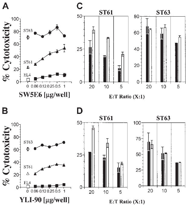
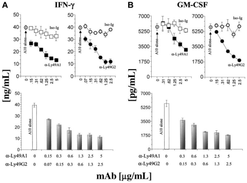
, anti-Ly49A and anti-Ly49G2 combined with indicated mAb concentrations.
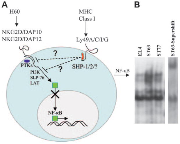
Figure 7
Figure 7. Inhibition of the NKG2D activation pathway is mediated through the reduced translocation of NF-κB into the nuclei of NK cells
(A) Diagram depicting the hypothesized interactions between activating NKG2D and inhibitory Ly49 pathways. Binding of H60 with NKG2D receptor leads to phosphorylation of DAP10 or DAP12, resulting in recruitment of protein tyrosine kinases (PTKs) belonging to the SFK family. The subsequent activation of PTKs results in the phosphorylation of downstream signaling proteins, eventually leading to nuclear translocation of NF-κB. Phosphorylation of ITIM sequences at the cytoplasmic domain of Ly49 molecules recruits and activates phosphatases, which in turn dephosphorylate substrates that are part of the NKG2D activation pathway. This results in the impaired translocation of NF-κB into the nuclei. (B) Gel-shift analysis was performed using nuclear extracts from fresh NK cells incubated with indicated target cells at a 40:1 E / T ratio for 2 hours at 37°C. Supershift analysis was performed on nuclear extracts from NK cells incubated with ST63 in the presence of 32P-labelled oligonucleotide and a mAb specific for NF-κB subunit, p50. Data presented are representative of 2 independent experiments.

Similar articles

Cited by

References

    1. Vyas YM, Maniar H, Dupont B. Visualization of signaling pathways and cortical cytoskeleton in cytolytic and noncytolytic natural killer cell immune synapses. Immunol Rev. 2002;189:161–178. - PubMed
    1. Karre K, Ljunggren HG, Piontek G, Kiessling R. Selective rejection of H-2-deficient lymphoma variants suggests alternative immune defence strategy. Nature. 1986;319:675–678. - PubMed
    1. Held W, Cado D, Raulet DH. Transgenic expression of the Ly49A natural killer cell receptor confers class I major histocompatibility complex (MHC)-specific inhibition and prevents bone marrow allograft rejection. J Exp Med. 1996;184:2037–2041. - PMC - PubMed
    1. Yu YY, George T, Dorfman JR, et al. The role of Ly49A and 5E6(Ly49C) molecules in hybrid resistance mediated by murine natural killer cells against normal T cell blasts. Immunity. 1996;4:67–76. - PubMed
    1. Hanke T, Takizawa H, McMahon CW, et al. Direct assessment of MHC class I binding by seven Ly49 inhibitory NK cell receptors. Immunity. 1999;11:67–77. - PubMed

Publication types

MeSH terms