. 2004 Aug 24;32(15):4539-49.
doi: 10.1093/nar/gkh778. Print 2004.

Increased frequency of aberrant V(D)J recombination products in core RAG-expressing mice

Affiliations

Increased frequency of aberrant V(D)J recombination products in core RAG-expressing mice

Sadiqur R Talukder et al. Nucleic Acids Res. .

Abstract

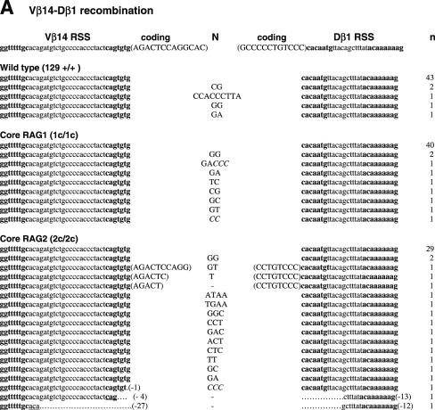
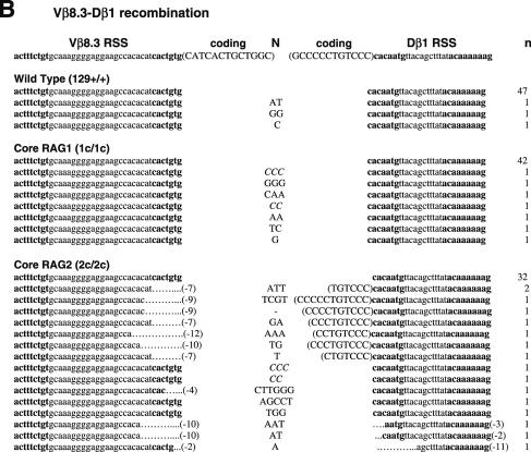
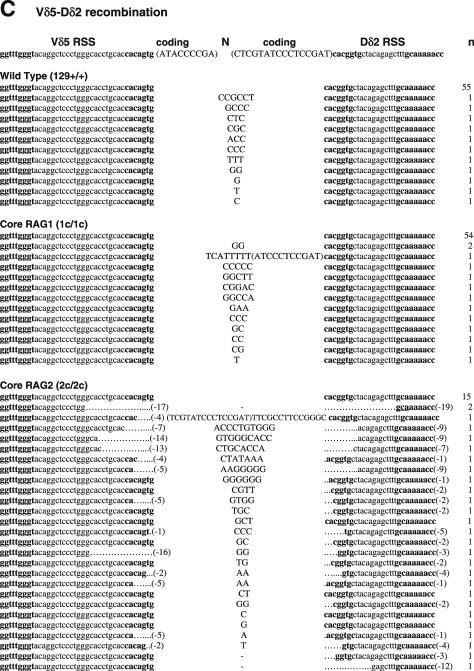
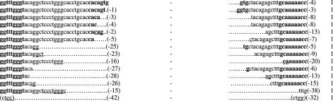
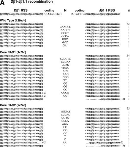
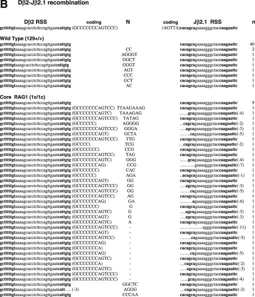
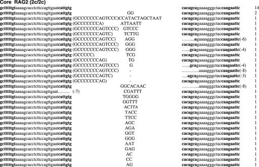
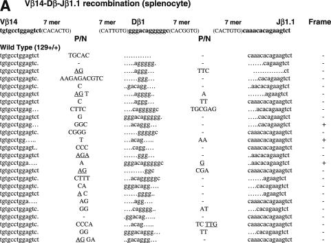
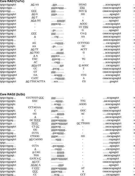
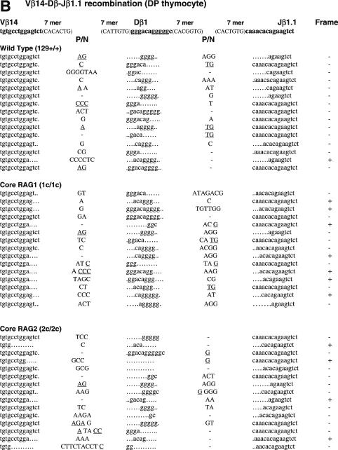
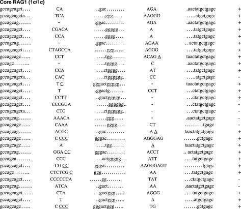
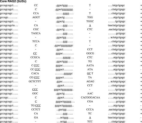
RAG1 and RAG2 play a central role in V(D)J recombination, a process for antigen receptor gene assembly. The truncated 'core' regions of RAGs are sufficient to catalyze the recombination reaction, although with lower joining efficiency than full-length proteins. To investigate the role of the non-core regions of RAGs in the end-joining phase of antigen receptor rearrangement, we analyzed recombination products isolated from core RAG1 and core RAG2 knock-in mice. Here, we report that the truncation of RAGs increases the frequency of aberrant recombination in vivo. Signal joints (SJs) associated with V-to-D recombination of core RAG1 knock-in mice were normal, whereas those of core RAG2 knock-in mice were highly imprecise, containing large deletions and additions, and in some cases coding sequences. In contrast, we found an elevated level of imprecise D-to-J associated SJs for both core RAG1- and RAG2-expressing mice. Likewise, sequences of coding joints (CJs) were also affected by the expression of core RAGs. Finally, sequences found at the junctions of rearranged T-cell receptor loci were highly influenced by differences in rearranging recombination signal sequence pairs. We provide the first evidence that the non-core regions of RAGs have critical functions in the proper assembly and resolution of recombination intermediates in endogenous antigen receptor loci.


Figures

Figure 1
Figure 1
Sequences of SJs formed by V-D recombination. (A) PCR fragments containing SJs formed by Vβ14-Dβ1 recombination were compared among WT (RAG1+/+ RAG2+/+), RAG1 core (RAG1c/c RAG2+/+) and RAG2 core (RAG1+/+ RAG2c/c) mice. The original sequences are indicated by lowercase letters and possible insertions are indicated in capital letters. Heptamer and nonamer sequences are in boldface letters. Nucleotide additions containing more than three continuous bases identical to either CEs were considered as coding sequence and indicated in parentheses. Nucleotide additions assigned as N additions with 2 or 3 nt homology with either CE are italicized. Numbers of deleted bases are shown in parentheses at the right end of each RSS. Microhomologies are underlined. ‘n’ indicates the number of junctions with the indicated sequence. (B) SJs formed by Vβ8.3-Dβ1 recombination. (C) SJ formed by Vδ5-Dδ2 recombination.
Figure 1
Figure 1
Sequences of SJs formed by V-D recombination. (A) PCR fragments containing SJs formed by Vβ14-Dβ1 recombination were compared among WT (RAG1+/+ RAG2+/+), RAG1 core (RAG1c/c RAG2+/+) and RAG2 core (RAG1+/+ RAG2c/c) mice. The original sequences are indicated by lowercase letters and possible insertions are indicated in capital letters. Heptamer and nonamer sequences are in boldface letters. Nucleotide additions containing more than three continuous bases identical to either CEs were considered as coding sequence and indicated in parentheses. Nucleotide additions assigned as N additions with 2 or 3 nt homology with either CE are italicized. Numbers of deleted bases are shown in parentheses at the right end of each RSS. Microhomologies are underlined. ‘n’ indicates the number of junctions with the indicated sequence. (B) SJs formed by Vβ8.3-Dβ1 recombination. (C) SJ formed by Vδ5-Dδ2 recombination.
Figure 2
Figure 2
Sequences of SJs formed by D-J recombination. (A) PCR fragments containing SJs formed by Dβ1-Jβ1.1 recombination were compared among WT (RAG1+/+ RAG2+/+), core RAG1 (RAG1c/c RAG2+/+) and core RAG2 (RAG1+/+ RAG2c/c) expressing mice. Details are as in Figure 1 legend. (B) SJ formed by Dβ2-Jβ2.1 recombination. (C) SJ formed by Dδ2-Jδ1 recombination.
Figure 2
Figure 2
Sequences of SJs formed by D-J recombination. (A) PCR fragments containing SJs formed by Dβ1-Jβ1.1 recombination were compared among WT (RAG1+/+ RAG2+/+), core RAG1 (RAG1c/c RAG2+/+) and core RAG2 (RAG1+/+ RAG2c/c) expressing mice. Details are as in Figure 1 legend. (B) SJ formed by Dβ2-Jβ2.1 recombination. (C) SJ formed by Dδ2-Jδ1 recombination.
Figure 3
Figure 3
Sequences of CJs formed by V-D-J recombination. (A) CJs formed by Vβ14-Dβ-Jβ1.1 recombination in splenocytes were compared among WT (RAG1+/+ RAG2+/+), core RAG1 (RAG1c/c RAG2+/+) and core RAG2 (RAG1+/+ RAG2c/c) expressing mice. In Vβ14-Dβ-Jβ1.1 recombination, Dβ1 within the Dβ1-Jβ1 gene cluster is expected to be utilized. Original coding sequences of Vβ14, Dβ1 and Jβ1.1 (boldface lowercase letters) and their flanking heptamer sequences (uppercase letters in parentheses) are indicated on the top. Nucleotide insertions are indicated by capital letters. Presumptive P nucleotides are underlined. All the clones contained different junctional sequences to each other. In-frame or out-of-frame rearrangement is denoted as ‘+’ or ‘−’ at the right end of each sequence. (B) CJ formed by Vβ14-Dβ-Jβ1.1 recombination in DP thymocytes. (C) CJ formed by Vβ10-Dβ-Jβ2.1 recombination in DP thymocytes. Original coding sequences of Vβ10, Dβ2 and Jβ2.1 (boldface lowercase letters) and their flanking heptamer sequences (uppercase letters in parentheses) are indicated on the top. Although Dβ1 can be used instead of Dβ2 in at least one-fourth of all Vβ10-Dβ-Jβ2.1 CJ (69), but they could not be assigned due to similarity. See (A) and (B) for Dβ1 coding sequence.
Figure 3
Figure 3
Sequences of CJs formed by V-D-J recombination. (A) CJs formed by Vβ14-Dβ-Jβ1.1 recombination in splenocytes were compared among WT (RAG1+/+ RAG2+/+), core RAG1 (RAG1c/c RAG2+/+) and core RAG2 (RAG1+/+ RAG2c/c) expressing mice. In Vβ14-Dβ-Jβ1.1 recombination, Dβ1 within the Dβ1-Jβ1 gene cluster is expected to be utilized. Original coding sequences of Vβ14, Dβ1 and Jβ1.1 (boldface lowercase letters) and their flanking heptamer sequences (uppercase letters in parentheses) are indicated on the top. Nucleotide insertions are indicated by capital letters. Presumptive P nucleotides are underlined. All the clones contained different junctional sequences to each other. In-frame or out-of-frame rearrangement is denoted as ‘+’ or ‘−’ at the right end of each sequence. (B) CJ formed by Vβ14-Dβ-Jβ1.1 recombination in DP thymocytes. (C) CJ formed by Vβ10-Dβ-Jβ2.1 recombination in DP thymocytes. Original coding sequences of Vβ10, Dβ2 and Jβ2.1 (boldface lowercase letters) and their flanking heptamer sequences (uppercase letters in parentheses) are indicated on the top. Although Dβ1 can be used instead of Dβ2 in at least one-fourth of all Vβ10-Dβ-Jβ2.1 CJ (69), but they could not be assigned due to similarity. See (A) and (B) for Dβ1 coding sequence.

Similar articles

Cited by

References

    1. Bassing C.H., Swat,W. and Alt,F.W. (2002) The mechanism and regulation of chromosomal V(D)J recombination. Cell, 109 (Suppl.), S45–S55. - PubMed
    1. Gellert M. (2002) V(D)J recombination: RAG proteins, repair factors, and regulation. Annu. Rev. Biochem., 71, 101–132. - PubMed
    1. Agrawal A. and Schatz,D.G. (1997) RAG1 and RAG2 form a stable postcleavage synaptic complex with DNA containing signal ends in V(D)J recombination. Cell, 89, 43–53. - PubMed
    1. Hiom K. and Gellert,M. (1998) Assembly of a 12/23 paired signal complex: a critical control point in V(D)J recombination. Mol. Cell, 1, 1011–1019. - PubMed
    1. Jones J.M. and Gellert,M. (2001) Intermediates in V(D)J recombination: a stable RAG1/2 complex sequesters cleaved RSS ends. Proc. Natl Acad. Sci. USA, 98, 12926–12931. - PMC - PubMed

Publication types