Review
. 2004 Aug 31;101(35):12783-8.
doi: 10.1073/pnas.0401586101. Epub 2004 Aug 24.

Multiple pathways process stalled replication forks

Affiliations
Review

Multiple pathways process stalled replication forks

Bénédicte Michel et al. Proc Natl Acad Sci U S A. .

Abstract

Impairment of replication fork progression is a serious threat to living organisms and a potential source of genome instability. Studies in prokaryotes have provided evidence that inactivated replication forks can restart by the reassembly of the replication machinery. Several strategies for the processing of inactivated replication forks before replisome reassembly have been described. Most of these require the action of recombination proteins, with different proteins being implicated, depending on the cause of fork arrest. The action of recombination proteins at blocked forks is not necessarily accompanied by a strand-exchange reaction and may prevent rather than repair fork breakage. These various restart pathways may reflect different structures at stalled forks. We review here the different strategies of fork processing elicited by different kinds of replication impairments in prokaryotes and the variety of roles played by recombination proteins in these processes.


Figures

Fig. 1.
Fig. 1.
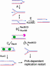
Replication fork reversal model (adapted from ref. 9). In the first step (A), the replication fork is arrested, causing fork reversal. The reversed fork forms an HJ (two alternative representations of this structure are shown, open X and parallel stacked X). In Rec+ cells (B and C), RecBCD initiates RecA-dependent homologous recombination at a chi site present on the DNA double-strand end, and the two HJ (one formed by reversal, one by homologous recombination) are resolved by RuvABC. Alternatively, if RecBCD encounters the HJ before encounter with chi or in the absence of RecA (B–D), the DNA double-strand end is degraded up to the HJ, restoring a fork structure. In both cases, replication restarts by a PriA-dependent process. In the absence of RecBCD (E), resolution of the HJ by RuvABC causes chromosome linearization. Continuous lines, parental chromosome; dashed lines, newly synthesized strands; green circle, RuvAB; pink incised circle, RecBCD.
Fig. 2.
Fig. 2.
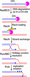
Recombination at a DNA double-strand end in bacteria. RecBCD binds to a DNA double-strand end and degrades both strands until it reaches a chi site. RecBCD loads RecA when it encounters chi and the RecA filament invades an homologous intact molecule. The HJ is resolved by RuvABC (or RecG). PriA-dependent primosome assembly promotes initiation of replication from the invading 3′ end. Blue and red lines, two homologous DNA molecules; pink incised circle, RecBCD; small yellow circles, RecA.
Fig. 3.
Fig. 3.
Model for RecA-dependent replication fork reversal (adapted from ref. 24). In the dnaBts recB strain, RuvABC-dependent chromosome linearization requires RecA. We propose that a RecA filament forms on the blocked lagging strand, which invades the homologous leading-strand resulting in a reversed replication fork. The HJ is then bound by RuvAB and the double-strand end by RecBCD. The processing of the reversed fork is as in Fig. 1. Green circle, RuvAB; small yellow circles, RecA.
Fig. 4.
Fig. 4.
Model for replication-dependent formation of linear DNA at Ter-blocked forks (adapted from ref. 34). Replication forks are arrested by ectopic Ter sites. Progression of a second round of replication forks up to the first blocked forks generates DNA double-strand ends. The linear DNA formed by overreplication will be repaired by homologous recombination (not shown). Black lines, parental chromosome; blue lines, first round of replication, blocked at Ter; red lines, strands of the second round of replication; O, chromosome origin; pink incised rectangle, Ter sites; yellow ovals, replisome.
Fig. 5.
Fig. 5.
Possible models for replication restart after UV irradiation. When a UV lesion forms downstream of a replication fork on the leading strand template, it may either block the entire replisome (A) or arrest the leading strand, whereas the lagging strand synthesis progresses further (B). In model A (adapted with modifications from ref. 44), RecFOR and RecA bind to the lagging strand template and promote the invasion of the leading strand, which renders the lesion double stranded and thus accessible to nucleotide excision repair (NER). We propose that the displaced leading strand end is degraded by a 3′ exonuclease (such as Exo I, Exo VII, or Exo X). In the absence of a functional NER system, or at very high UV doses saturating NER, bypass polymerases would be needed to incorporate a nucleotide opposite the lesion site. In model B (adapted with modifications from ref. 6), the lagging strand progresses further than the leading strand. In contrast with model A, both lagging and leading strand ends need to be displaced to render the lesion double-stranded, hence accessible to NER, resulting in a reversed fork. The lesion could be repaired either before or after (drawn here) fork resetting. The question marks indicate that, considering the lack of requirement for RecBC, RuvABC, and/or RecG for replication restart after UV irradiation, how the reversed fork would be formed and reset is presently unknown. Continuous lines, parental chromosome; dashed lines, newly synthesized strands; small yellow circles, RecA; pink rectangle, RecFOR; black triangle, DNA lesion.

References

    1. Kogoma, T. (1997) Microbiol. Mol. Biol. Rev. 61, 212–238. - PMC - PubMed
    1. Sandler, S. J. & Marians, K. J. (2000) J. Bacteriol. 182, 9–13. - PMC - PubMed
    1. Xu, L. & Marians, K. J. (2003) Mol. Cell 11, 817–826. - PubMed
    1. Kraus, E., Leung, W. Y. & Haber, J. E. (2001) Proc. Natl. Acad. Sci. USA 98, 8255–8262. - PMC - PubMed
    1. Ira, G. & Haber, J. E. (2002) Mol. Cell. Biol. 22, 6384–6392. - PMC - PubMed

LinkOut - more resources