

A novel 1p36.2 located gene, APITD1, with tumour-suppressive properties and a putative p53-binding domain, shows low expression in neuroblastoma tumours
- PMID: 15328517
- PMCID: PMC2747717
- DOI: 10.1038/sj.bjc.6602083
A novel 1p36.2 located gene, APITD1, with tumour-suppressive properties and a putative p53-binding domain, shows low expression in neuroblastoma tumours
Abstract
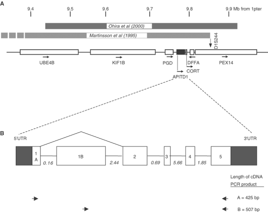
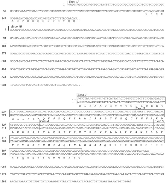
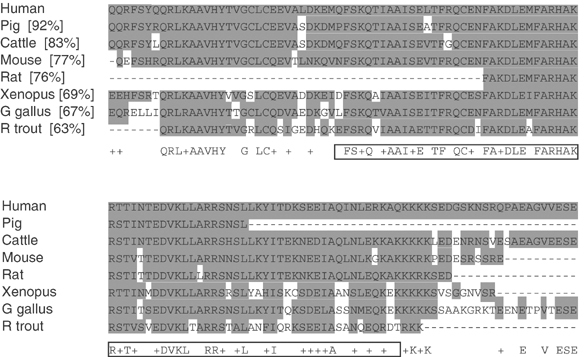
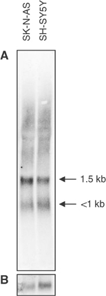
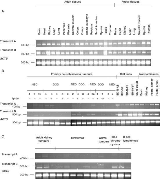
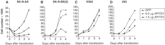
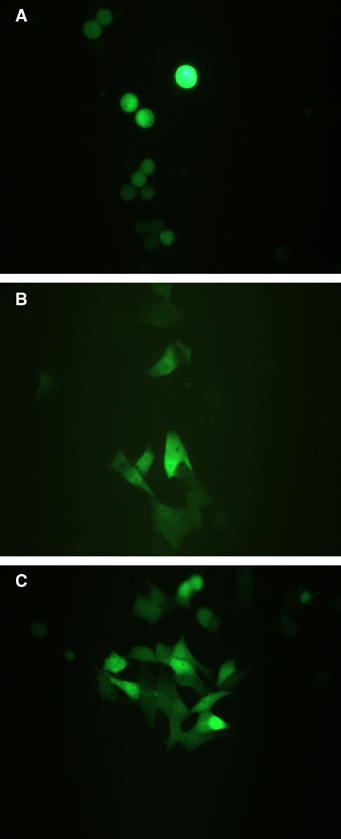
Neuroblastoma is characterised by a lack of TP53 mutations and no other tumour suppressor gene consistently inactivated has yet been identified in this childhood cancer form. Characterisation of a new gene, denoted APITD1, in the neuroblastoma tumour suppressor candidate region in chromosome 1p36.22 reveals that APITD1 contains a predicted TFIID-31 domain, representing the TATA box-binding protein-associated factor, TAF(II)31, which is required for p53-mediated transcription activation. Two different transcripts of this gene were shown to be ubiquitously expressed, one of them with an elevated expression in foetal tissues. Primary neuroblastoma tumours of all different stages showed either very weak or no measurable APITD1 expression, contrary to the level of expression observed in neuroblastoma cell lines. A reduced pattern of expression was also observed in a set of various tumour types. APITD1 was functionally tested by adding APITD1 mRNA to neuroblastoma cells, leading to the cell growth to be reduced up to 90% compared to control cells, suggesting APITD1 to have a role in a cell death pathway. Furthermore, we determined the genomic organisation of APITD1. Automated genomic DNA sequencing of the coding region of the gene as well as the promoter sequence in 44 neuroblastoma tumours did not reveal any loss-of-function mutations, indicating that mutations in APITD1 is not a common abnormality of neuroblastoma tumours. We suggest that low expression of this gene might interfere with the ability for apoptosis through the p53 pathway.
Figures
Similar articles
-
Expression of APITD1 is not related to copy number changes of chromosomal region 1p36 or the prognosis of uveal melanoma.Invest Ophthalmol Vis Sci. 2007 Nov;48(11):4919-23. doi: 10.1167/iovs.07-0061. Invest Ophthalmol Vis Sci. 2007. PMID: 17962439
-
A cluster of genes located in 1p36 are down-regulated in neuroblastomas with poor prognosis, but not due to CpG island methylation.Mol Cancer. 2005 Mar 1;4(1):10. doi: 10.1186/1476-4598-4-10. Mol Cancer. 2005. PMID: 15740626 Free PMC article.
-
Analyses of apoptotic regulators CASP9 and DFFA at 1P36.2, reveal rare allele variants in human neuroblastoma tumours.Br J Cancer. 2002 Feb 12;86(4):596-604. doi: 10.1038/sj.bjc.6600111. Br J Cancer. 2002. PMID: 11870543 Free PMC article.
-
[Identification of the breakpoint-flanking markers on chromosomes 1 and 17 of a constitutional translocation T(1;17)(P36;Q12-21) in a patient with neuroblastoma].Verh K Acad Geneeskd Belg. 1995;57(5):389-422. Verh K Acad Geneeskd Belg. 1995. PMID: 8571670 Review. Dutch.
-
The quest for the 1p36 tumor suppressor.Cancer Res. 2008 Apr 15;68(8):2551-6. doi: 10.1158/0008-5472.CAN-07-2095. Cancer Res. 2008. PMID: 18413720 Free PMC article. Review.
Cited by
-
Fine map of the Gct1 spontaneous ovarian granulosa cell tumor locus.Mamm Genome. 2013 Feb;24(1-2):63-71. doi: 10.1007/s00335-012-9439-6. Epub 2012 Nov 18. Mamm Genome. 2013. PMID: 23179634 Free PMC article.
-
Characterization of a FOXG1:TLE1 transcriptional network in glioblastoma-initiating cells.Mol Oncol. 2018 Jun;12(6):775-787. doi: 10.1002/1878-0261.12168. Epub 2018 Apr 27. Mol Oncol. 2018. PMID: 29316219 Free PMC article.
-
hsa_circ_0077837 Alleviated the Malignancy of Non-Small Cell Lung Cancer by Regulating the miR-1178-3p/APITD1 Axis.J Oncol. 2022 Mar 9;2022:3902832. doi: 10.1155/2022/3902832. eCollection 2022. J Oncol. 2022. PMID: 35310916 Free PMC article.
-
A Numerically Subdominant CD8 T Cell Response to Matrix Protein of Respiratory Syncytial Virus Controls Infection with Limited Immunopathology.PLoS Pathog. 2016 Mar 4;12(3):e1005486. doi: 10.1371/journal.ppat.1005486. eCollection 2016 Mar. PLoS Pathog. 2016. PMID: 26943673 Free PMC article.
-
A histone-fold complex and FANCM form a conserved DNA-remodeling complex to maintain genome stability.Mol Cell. 2010 Mar 26;37(6):865-78. doi: 10.1016/j.molcel.2010.01.039. Mol Cell. 2010. PMID: 20347428 Free PMC article.
References
-
- Bauer A, Savelyeva L, Claas A, Praml C, Berthold F, Schwab M (2001) Smallest region of overlapping deletion in 1p36 in human neuroblastoma: a 1 Mbp cosmid and PAC contig. Genes Chromosomes Cancer 31: 228–239 - PubMed
-
- Benn DE, Dwight T, Richardson AL, Delbridge L, Bambach CP, Stowasser M, Gordon RD, Marsh DJ, Robinson BG (2000) Sporadic and familial pheochromocytomas are associated with loss of at least two discrete intervals on chromosome 1p. Cancer Res 60: 7048–7051 - PubMed
-
- Bridge JA, Liu J, Weibolt V, Baker KS, Perry D, Kruger R, Qualman S, Barr F, Sorensen P, Triche T, Suijkerbuijk R (2000) Novel genomic imbalances in embryonal rhabdomyosarcoma revealed by comparative genomic hybridization and fluorescence in situ hybridization: an intergroup rhabdomyosarcoma study. Genes Chromosomes Cancer 27: 337–344 - PubMed
-
- Brodeur GM, Green AA, Hayes FA, Williams KJ, Williams DL, Tsiatis AA (1981) Cytogenetic features of human neuroblastomas and cell lines. Cancer Res 41: 4678–4686 - PubMed
Publication types
MeSH terms
Substances
LinkOut - more resources
Full Text Sources
Medical
Molecular Biology Databases
Research Materials
Miscellaneous
