

METH-2 silencing and promoter hypermethylation in NSCLC
- PMID: 15328519
- PMCID: PMC2747718
- DOI: 10.1038/sj.bjc.6602107
METH-2 silencing and promoter hypermethylation in NSCLC
Abstract
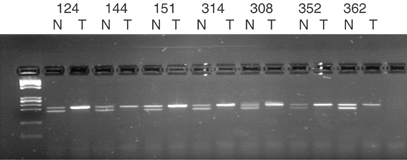
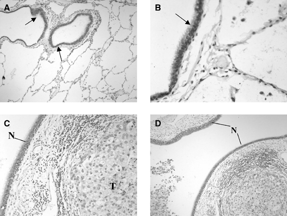
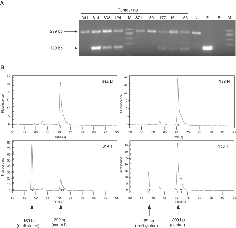
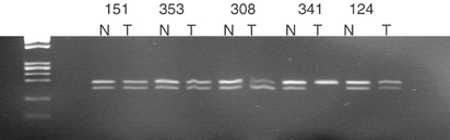
The antiangiogenic factor METH-2 (ADAMTS-8) was identified in a previous dual-channel cDNA microarray analysis to be at least two-fold under-represented in 85% (28 out of 33) of primary non-small-cell lung carcinomas (NSCLCs). This observation has been validated in an independent series of NSCLCs and adjacent normal tissues by comparative multiplex RT-PCR, and METH-2 mRNA expression was dramatically reduced in all 23 tumour samples analysed. Immunohistochemical analysis of the same sample set demonstrated that METH-2 was strongly expressed in 14 out of 19 normal epithelial sites examined but only one out of 20 NSCLCs. DNA methylation analysis of the proximal promoter region of this gene revealed abnormal hypermethylation in 67% of the adenocarcinomas and 50% of squamous cell carcinomas, indicating that epigenetic mechanisms are involved in silencing this gene in NSCLC. No homozygous deletions of METH-2 were found in lung cancer cell lines. Allelic imbalance in METH-2 was assessed by an intronic single nucleotide polymorphism (SNP) assay and observed in 44% of informative primary samples. In conclusion, the downregulation of METH-2 expression in primary NSCLC, often associated with promoter hypermethylation, is a frequent event, which may be related to the development of the disease.
Figures
References
-
- Cox G, Jones JL, Walker RA, Steward WP, O'Byrne KJ (2000) Angiogenesis and non-small cell lung cancer. Lung Cancer 27: 81–100 - PubMed
-
- Folkman J, Cotran R (1976) Relation of vascular proliferation to tumor growth. Int Rev Exp Pathol 16: 207–248 - PubMed
-
- Folkman J, Watson K, Ingber D, Hanahan D (1989) Induction of angiogenesis during the transition from hyperplasia to neoplasia. Nature 339: 58–61 - PubMed
-
- Georgiadis KE, Hirohata S, Seldin MF, Apte SS (1999) ADAM-TS8, a novel metalloprotease of the ADAM-TS family located on mouse chromosome 9 and human chromosome 11. Genomics 62: 312–315 - PubMed
-
- Heighway J, Betticher D, Knapp T, Hoban P (2003) Comparative multiplex PCR and allele-specific expression analysis in human lung cancer. Tools to facilitate target identification. Methods Mol Med 75: 291–304 - PubMed
Publication types
MeSH terms
Substances
LinkOut - more resources
Full Text Sources
Medical
Molecular Biology Databases
