

Induction of apoptosis in human prostate cancer cell line, PC3, by 3,3'-diindolylmethane through the mitochondrial pathway
- PMID: 15328526
- PMCID: PMC2409910
- DOI: 10.1038/sj.bjc.6602145
Induction of apoptosis in human prostate cancer cell line, PC3, by 3,3'-diindolylmethane through the mitochondrial pathway
Abstract
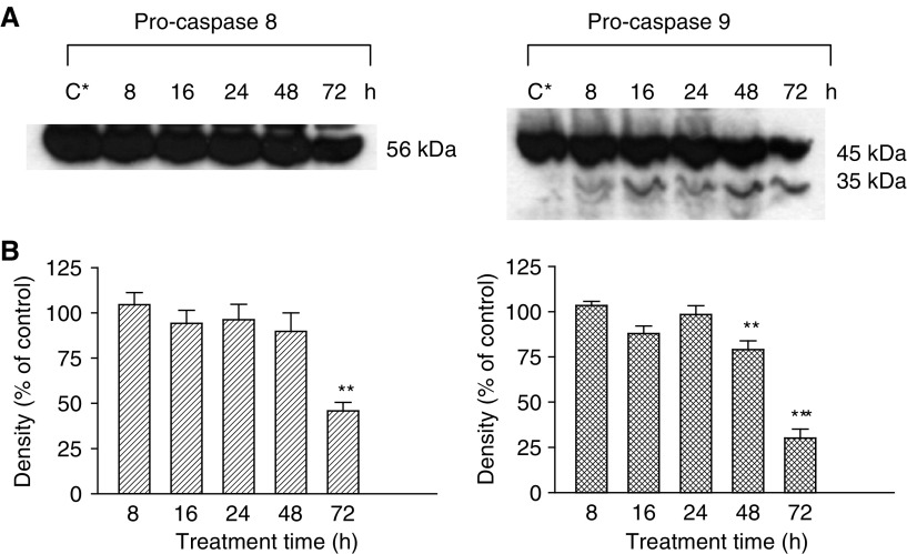
Prostate cancer is the most common malignancy and the second leading cause of male death in Western countries. Prostate cancer mortality results from metastases to the bones and lymph nodes and progression from androgen-dependent to androgen-independent disease. Although androgen ablation was found to be effective in treating androgen-dependent prostate cancer, no effective life-prolonging therapy is available for androgen-independent cancer. Epidemiological studies have shown a strong correlation between consumption of cruciferous vegetables and a lower risk of prostate cancer. These vegetables contain glucosinolates, which during metabolism give rise to several breakdown products, mainly indole-3-carbinol (I3C), which may be condensed to polymeric products, especially 3,3'-diindolylmethane (DIM). It was previously shown that these indole derivatives have significant inhibitory effects in several human cancer cell lines, which are exerted through induction of apoptosis. We have previously reported that I3C and DIM induce apoptosis in prostate cancer cell lines through p53-, bax-, bcl-2- and fasL-independent pathways. The objective of this study was examination of the apoptotic pathways that may be involved in the effect of DIM in the androgen-independent prostate cancer cell line, PC3, in vitro. Our results suggest that DIM induces apoptosis in PC3 cells, through the mitochondrial pathway, which involves the translocation of cytochrome c from the mitochondria to the cytosol and the activation of initiator caspase, 9, and effector caspases, 3 and 6, leading to poly ADP-ribose polymerase (PARP) cleavage and induction of apoptosis. Our findings may lead to the development of new therapeutic strategies for the treatment of androgen-independent prostate cancer.
Figures


References
-
- Abate-Shen C, Shen MM (2000) Molecular genetics of prostate cancer. Genes Dev 14: 2410–2434 - PubMed
-
- Bonnesen C, Eggleston IM, Hayes JD (2001) Dietary indoles and isothiocyanates that are generated from cruciferous vegetables can both stimulate apoptosis and confer protection against DNA damage in human colon cell lines. Cancer Res 61: 6120–6130 - PubMed
-
- Bortner CD, Oldenburg NBE, Cidlowski JA (1995) The role of DNA fragmentation in apoptosis. Trends Cell Biol 5: 21–26 - PubMed
-
- Bradfield CA, Bjeldanes LF (1987) High-performance liquid chromatographic analysis of anticarcinogenic indoles in Brassica oleracea. J Agric Food Chem 35: 46–49
MeSH terms
Substances
LinkOut - more resources
Full Text Sources
Medical
Research Materials
Miscellaneous
