. 2004 Sep;2(9):E274.
doi: 10.1371/journal.pbio.0020274. Epub 2004 Aug 24.

Recognition and accommodation at the androgen receptor coactivator binding interface

Affiliations

Recognition and accommodation at the androgen receptor coactivator binding interface

Eugene Hur et al. PLoS Biol. 2004 Sep.

Abstract

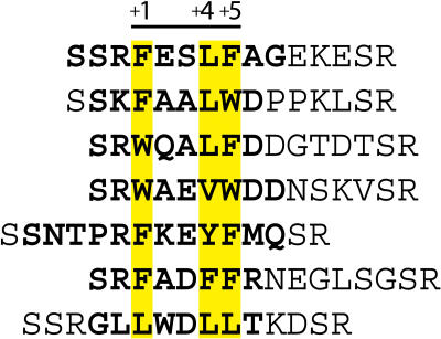
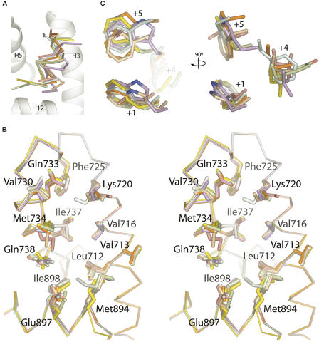
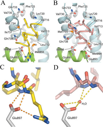
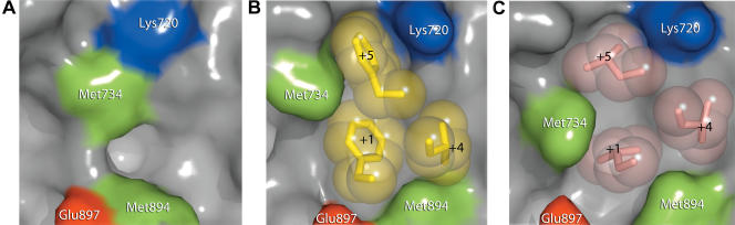
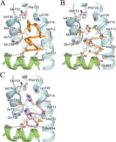
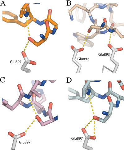
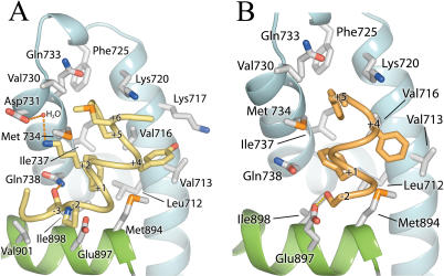
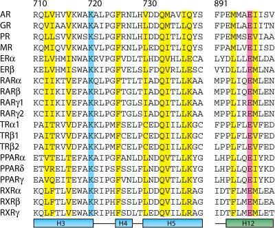
Prostate cancer is a leading killer of men in the industrialized world. Underlying this disease is the aberrant action of the androgen receptor (AR). AR is distinguished from other nuclear receptors in that after hormone binding, it preferentially responds to a specialized set of coactivators bearing aromatic-rich motifs, while responding poorly to coactivators bearing the leucine-rich "NR box" motifs favored by other nuclear receptors. Under normal conditions, interactions with these AR-specific coactivators through aromatic-rich motifs underlie targeted gene transcription. However, during prostate cancer, abnormal association with such coactivators, as well as with coactivators containing canonical leucine-rich motifs, promotes disease progression. To understand the paradox of this unusual selectivity, we have derived a complete set of peptide motifs that interact with AR using phage display. Binding affinities were measured for a selected set of these peptides and their interactions with AR determined by X-ray crystallography. Structures of AR in complex with FxxLF, LxxLL, FxxLW, WxxLF, WxxVW, FxxFF, and FxxYF motifs reveal a changing surface of the AR coactivator binding interface that permits accommodation of both AR-specific aromatic-rich motifs and canonical leucine-rich motifs. Induced fit provides perfect mating of the motifs representing the known family of AR coactivators and suggests a framework for the design of AR coactivator antagonists.


Conflict of interest statement

The authors have declared that no conflicts of interest exist.

Figures

Figure 1
Figure 1. AR LBD–Interacting Peptides Selected by Phage Display
Hydrophobic residues of the core motif are highlighted in yellow. Residues in bold were ordered in electron density maps.
Figure 2
Figure 2. A Structural Profile of the AR Coactivator Binding Interface
AR–peptide complexes are colored as follows: FxxLF, yellow; FxxLW, orange; WxxLF, wheat; WxxVW, purple; FxxYF, green; FxxFF, blue; LxxLL, pink; unbound, grey. (A) Cα trace of the peptides superimposed onto the AF-2. For clarity only the LBD of AR–FxxLF is shown. (B) Superposition of the LBD of the AR–peptide complexes in the region of the coactivator interface. Backbone atoms are shown as a Cα trace. Side chains of residues composing the interface are shown as sticks. (C) Hydrophobic side chains of the core motif superimposed as in (B).
Figure 3
Figure 3. Interactions of FxxLF and LxxLL with the AR LBD
(A and B) FxxLF (A) and LxxLL (B) bound to the AR AF-2 interface. FxxLF and LxxLL are shown as yellow and pink Cα coils, respectively. Helices 3, 4, and 5 of the LBD are shown as blue ribbons; Helix 12 is shown in green. LBD residues interacting with peptides are depicted as white sticks. For clarity only peptide side chains making significant interactions with the LBD are shown. (C and D) Hydrogen-bonding interactions between backbone atoms of FxxLF (C) and LxxLL (D) with Glu897 of the LBD. Peptide alpha carbons are labeled.
Figure 4
Figure 4. Induced Fit of the AR AF-2 Interface
Surface representations of the AR AF-2 interface. The unbound structure is shown in (A), the FxxLF bound in (B), and the LxxLL bound in (C). Side chains of the hydrophobic residues of the core motifs of FxxLF and LxxLL are shown as spheres.
Figure 5
Figure 5. Interactions of the Tryptophan Motifs with the AR LBD
FxxLW (A), WxxLF (B), and WxxVW (C) bound to the AR AF-2 interface. FxxLW, WxxLF, and WxxVW are shown as orange, beige, and purple Cα coils, respectively. The LBD is depicted as in Figure 3.
Figure 6
Figure 6. Interactions of Ser−2 with Glu897
Interactions between Ser−2 of the peptides (A) FxxLW, (B) WxxLF, (C) WxxVW, and (D) FxxFF and Glu897 of the LBD. Peptide alpha carbons are labeled.
Figure 7
Figure 7. Interactions of FxxYF and FxxFF with the AR LBD
FxxYF (A) and FxxFF (B) bound to the AR AF-2 interface. FxxYF and FxxFF are shown as yellow and orange Cα coils, respectively. The LBD is depicted as in Figure 3.
Figure 8
Figure 8. Sequence Alignment of the AF-2 Region of NRs
Residues composing the coactivator interface of AR are highlighted in yellow. The absolutely conserved glutamate and lysine composing the charge clamp are highlighted in pink and blue, respectively. Residue numbering is that of AR.
Figure 9
Figure 9. Surface Complimentarity of Hydrophobic Motifs in the AR, ERα, and GR AF-2 Clefts
(A) AR–FxxLF, (B)AR–LxxLL, (C) ERα–GRIP1 (LxxLL) (Shiau et al. 1998), and (D) GR-TIF2 (LxxLL) (Bledsoe et al. 2002). The inside surfaces of the AF-2 cleft in AR, ERα, and GR are depicted. The LBD is additionally shown as a Cα trace with key side chains shown as white sticks. Phenylalanines and leucines of the FxxLF and LxxLL motifs are shown as spheres.

References

    1. Alen P, Claessens F, Verhoeven G, Rombauts W, Peeters B. The androgen receptor amino-terminal domain plays a key role in p160 coactivator-stimulated gene transcription. Mol Cell Biol. 1999;19:6085–6097. - PMC - PubMed
    1. Asada S, Choi Y, Uesugi M. A gene-expression inhibitor that targets an alpha-helix-mediated protein interaction. J Am Chem Soc. 2003;125:4992–4993. - PubMed
    1. Bledsoe RK, Montana VG, Stanley TB, Delves CJ, Apolito CJ, et al. Crystal structure of the glucocorticoid receptor ligand binding domain reveals a novel mode of receptor dimerization and coactivator recognition. Cell. 2002;110:93–105. - PubMed
    1. Brunger AT, Adams PD, Clore GM, DeLano WL, Gros P, et al. Crystallography & NMR system: A new software suite for macromolecular structure determination. Acta Crystallogr D Biol Crystallogr. 1998;54:905–921. - PubMed
    1. Chang C, Norris JD, Gron H, Paige LA, Hamilton PT, et al. Dissection of the LXXLL nuclear receptor-coactivator interaction motif using combinatorial peptide libraries: Discovery of peptide antagonists of estrogen receptors alpha and beta. Mol Cell Biol. 1999;19:8226–8239. - PMC - PubMed

Publication types

MeSH terms

Substances