Comparative Study
. 2004 Sep;45(9):1091-9.
doi: 10.1111/j.0013-9580.2004.65803.x.

Is intraoperative electrocorticography reliable in children with intractable neocortical epilepsy?

Affiliations
Comparative Study

Is intraoperative electrocorticography reliable in children with intractable neocortical epilepsy?

Eishi Asano et al. Epilepsia. 2004 Sep.

Abstract

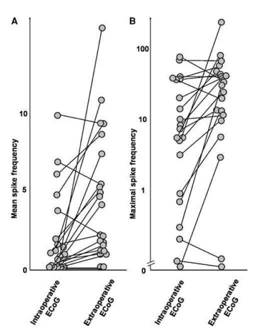
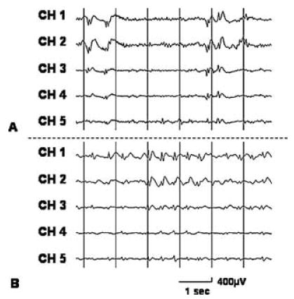
Purpose: To study the relation between the spike frequency during intraoperative electrocorticography (ECoG) under general anesthesia with isoflurane and that during extraoperative ECoG monitoring in children with intractable neocortical epilepsy.

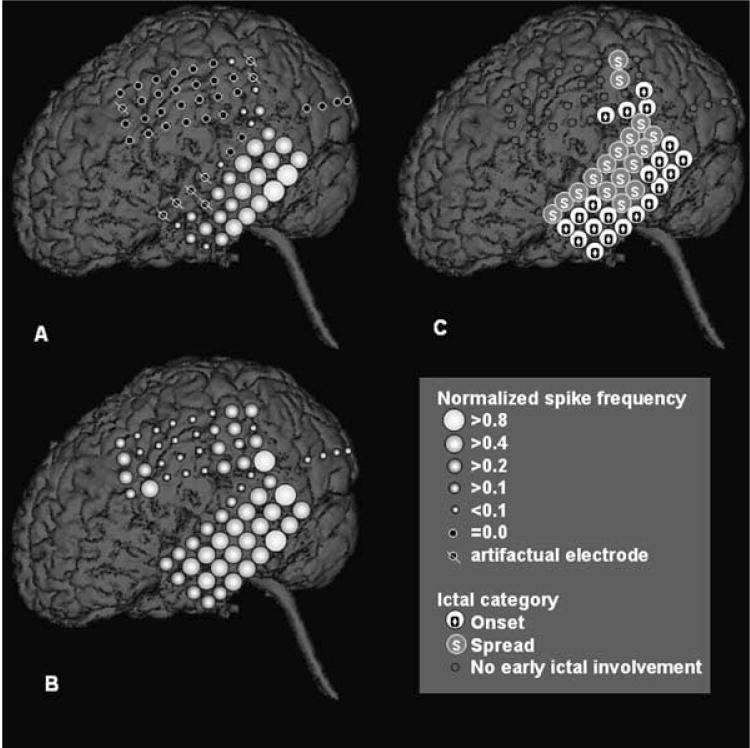
Methods: Twenty-one children (age, 1-16 years; 15 boys and six girls) who underwent intraoperative and extraoperative ECoG monitoring with subdural electrode arrays were studied. The spike frequency and the spatial pattern of spike frequency were compared between intraoperative and extraoperative ECoGs for each patient (by using Wilcoxon signed-ranks and Spearman's rank correlation, respectively).

Results: In 15 of 21 patients, the spike frequency was significantly lower during intraoperative than during extraoperative ECoG (mean z = -6.3; p < 0.001). In four of 21 patients, no significant difference was found in the spike frequency between intraoperative and extraoperative recordings. In two of 21 patients, spike frequency reached one spike/min neither during intraoperative nor extraoperative recording; therefore appropriate comparison of spike frequency was not possible. A significant positive correlation in the spike-frequency pattern was seen between intraoperative and extraoperative recordings in nine of nine cases who had > or = 10 spikes/min during intraoperative ECoG (mean rho = 0.62; p < 0.01), in five of six cases with one to nine spikes/min (mean rho = 0.50; p < 0.01), and in none of five cases with less than one spike/min (mean rho = 0.13).

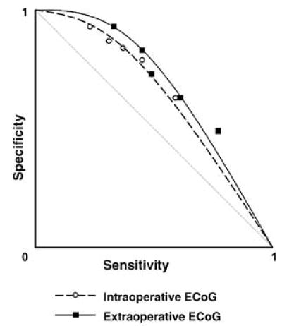
Conclusions: General anesthesia often decreases the spike frequency in children with neocortical epilepsy, yet intraoperative ECoG can reliably reflect the awake interictal spiking pattern when spike frequency exceeds one spike/min during intraoperative ECoG recording.


Figures

FIG. 1
FIG. 1
Interictal spike frequency during intraoperative and extra-operative electrocorticography recording in 21 children with neo-cortical epilepsy. Each circle represents mean spike frequency (A) and maximal spike frequency (B) for each patient.
FIG. 2
FIG. 2
A 5-year-old boy with a diagnosis of tuberous sclerosis complex and uncontrolled seizures. A: Normalized spike frequency during intraoperative electrocorticography (ECoG) is shown. The maximal spike frequency (8.3 spikes/min) was seen in the left posterior temporal region. B: Normalized spike frequency during extraoperative ECoG is shown. The maximal spike frequency (37.4 spikes/min) was also seen in the posterior temporal region. Although the left frontal region also showed frequent interictal spike activity, none of the frontal spikes appeared independent of the spikes in the left temporal region. Both spike topography mappings showed a similar gradient of spike frequency, being highest in the posterior temporal region, intermediate in the anterior temporal region and the parietal region, and lowest in the primary sensorimotor cortex and the lateral occipital region. The Spearman’s rank correlation demonstrated a significant positive correlation in the spike-frequency pattern between intraoperative and extraoperative ECoG recordings (rho = 0.62; p < 0.001). C: Ictal ECoG data are shown. Ictal onset involved the left temporal neocortex and a portion of the inferior parietal region. Ictal discharges propagated to the entire temporal region and a portion of the parietal region within 10 s from the seizure onset.
FIG. 3
FIG. 3
Independent repetitive spike–wave bursts on intraoperative and extraoperative electrocorticography (ECoG) in a 10-year-old girl with uncontrolled seizures. Both (A) intraoperative and (B) extraoperative ECoG showed frequent independent spike–wave bursts in the right subfrontal region. Ictal ECoG showed rhythmic fast-wave bursts in the right middle and superior frontal regions, and the subfrontal regions did not receive early ictal propagation. Brain magnetic resonance imaging showed a subcortical hetero-topia in the right subfrontal region, and glucose positron emission tomography scan showed glucose hypometabolism in the cortex overlying the heterotopia.
FIG. 4
FIG. 4
Receiver operating characteristics (ROC) curve. Cutoff thresholds of 40%, 20%, 15%, 10%, and 5% of the maximal spike frequency during intraoperative electrocorticography (ECoG) resulted in mean sensitivity of 0.22, 0.30, 0.35, 0.44, and 0.57, as well as mean specificity of 0.94, 0.87, 0.85, 0.79, and 0.63 for detection of “onset” electrodes, respectively. Similarly, cutoff thresholds of 40%, 20%, 15%, 10%, and 5% of the maximal spike frequency during extraoperative ECoG resulted in mean sensitivity of 0.32, 0.45, 0.47, 0.59, and 0.75, as well as mean specificity of 0.93, 0.84, 0.74, 0.64, and 0.50 for detection of “onset” electrodes. Sensitivity and specificity for each cutoff threshold are shown by white circles (for intraoperative ECoG) and black squares (for extraoperative ECoG).

References

    1. Luders HO, Engel J, Jr, Munari C. General Principles. In: Engel J Jr, editor. Surgical treatment of epilepsies. 1993. 2nd ed. New York: Raven Press; pp. 137–53.
    1. Keene DL, Whiting S, Ventureyra EC. Electrocorticography. Epileptic Disord. 2000;2:57–63. - PubMed
    1. Onal C, Otsubo H, Araki T, et al. Complications of invasive subdural grid monitoring in children with epilepsy. J Neurosurg. 2003;98:1017–26. - PubMed
    1. Fiol ME, Boening JA, Cruz-Rodriguez R, et al. Effect of isoflurane (Forane) on intraoperative electrocorticogram. Epilepsia. 1993;34:897–900. - PubMed
    1. Ito BM, Sato S, Kufta CV, et al. Effect of isoflurane and enflurane on the electrocorticogram of epileptic patients. Neurology. 1988;38:924–8. - PubMed

Publication types

MeSH terms