. 2004 Aug 25;24(34):7549-56.
doi: 10.1523/JNEUROSCI.1871-04.2004.

Substance P-mediated modulation of pacemaker properties in the mammalian respiratory network

Affiliations

Substance P-mediated modulation of pacemaker properties in the mammalian respiratory network

Fernando Peña et al. J Neurosci. .

Abstract

Neuromodulators are integral parts of a neuronal network, and unraveling how these substances alter neuronal activity is critical for understanding how networks generate patterned activity and, ultimately, behavior. In this study, we examined the cellular mechanisms underlying the excitatory action of substance P (SP) on the respiratory network isolated in spontaneously active transverse slice preparation of mice. SP produced a slow depolarization in all recorded inspiratory pacemaker and non-pacemaker neurons. Ion exchange experiments and blockers for different ion channels suggest that the slow depolarization is caused by the activation of a low-threshold TTX-insensitive cationic current that carries mostly Na+. The SP-induced slow depolarization increased tonic discharge in non-pacemaker neurons and primarily enhanced the frequency of bursting in Cd2+-insensitive pacemaker neurons. In the Cd2+-sensitive pacemaker neuron, the burst frequency was not significantly affected, whereas burst duration and amplitude were more enhanced than in Cd2+-insensitive pacemaker neurons. In a subset of non-pacemaker neurons that produced NMDA-dependent subthreshold oscillations, SP caused the production of bursts of action potentials. We conclude that the degree of pacemaker activity in the respiratory network is not fixed but dynamically regulated by neuromodulators such as SP. This finding may have clinical implications for Rett syndrome in which SP levels along with other neuromodulators are decreased in the brainstem.


Figures
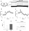
Figure 1.
Figure 1.
SP excites the respiratory network and depolarizes respiratory neurons. A, Schematic showing anatomical landmarks of a slice from the neonatal mouse medulla and recording sites for both population activity [integrated VRG (∫VRG)] and whole-cell patch-clamp recordings [membrane potential recording (VM)]. Note that application of 0.75 μm SP induces an excitation of integrated population activity (top trace) that is accompanied by a slow depolarization of a rhythmic neuron (bottom trace). The constant hyperpolarizing pulse was applied to test any change in input resistance produced by the application of SP. IO, Inferior olive; NA, nucleus ambiguus; SP5, spinally projecting trigeminal nucleus; XII, hypoglossal nucleus. B, Time course of the effect of SP on integrated population burst frequency. After 3 min of control recording, 0.75 μm SP was added (arrow) and continued for the rest of the experiment (n = 13). C, Time course of the effect of SP on the irregularity score (see Material and Methods) of respiratory activity. Note that after application of SP (arrow), the irregularity score is considerably reduced, indicating that the rhythm became more regular (n = 13). D, Quantification of resting membrane potential before [control (CON)] and after SP application for 15 respiratory neurons showing a significant depolarization. The asterisk denotes a statistically significant difference (p < 0.05). E, Comparison of a constant hyperpolarizing pulse as in A before (Control) and during application of SP, showing that there is no change on input resistance accompanying the depolarization induced by SP.
Figure 2.
Figure 2.
The SP-induced slow depolarization does not require synaptic transmission. A, Application of a mixture containing 10 μm CPP, 20 μm CNQX, 1 μm strychnine, and 20 μm bicuculline blocks synaptic transmission and the respiratory rhythm (∫VRG). Under these conditions, SP produces the same slow depolarization compared with the situation in the functional network (compare with Fig. 1A). The neuron presented was previously confirmed as a rhythmic respiratory neuron in the absence of the mixture. B, The SP-induced slow depolarization is accompanied by an increase in excitability. In the presence of SP, the same current injection produced more action potentials and an additional depolarizing drive potential. C, Quantification of resting membrane potential in the presence of the mixture before [control (CON)] and after SP application for 39 respiratory neurons shows a significant depolarization. The asterisk denotes a statistically significant difference (p < 0.05).
Figure 3.
Figure 3.
SP potentiates Cd2+-sensitive pacemaker activity. A, Example of a Cd2+-sensitive pacemaker neuron that continues to burst rhythmically (bottom trace) in the absence of rhythmic population activity (top trace). The recording was obtained in the presence of the mixture. The SP-induced slow depolarization is accompanied by a dramatic increase in burst generation leading to action potential inactivation. Note also the amplitude of bursting was affected during the peak depolarization, suggesting also the inactivation of the burst mechanisms themselves. Injection of the constant negative DC current (arrow) restores burst and action potential generation. The neuron presented was previously confirmed as an inspiratory neuron in the absence of the mixture. B, Same pacemaker neuron as A in an expanded timescale showing burst activity before (left), at the beginning of the SP-induced slow depolarization before action potential inactivation (middle), and after applying negative DC current to restore action potential activity (right). C-E, Histograms characterizing bursting properties of Cd2+-sensitive pacemaker neurons before [control (CON)] and during SP application. The asterisk denotes a statistically significant difference (p < 0.05).
Figure 4.
Figure 4.
SP excites Cd2+-insensitive pacemaker activity. A, Example of a Cd2+-insensitive pacemaker neuron that continues to burst rhythmically (bottom trace) in the absence of rhythmic population activity (top trace). The SP-induced slow depolarization increases bursting in this type of pacemaker. B, Same pacemaker neuron as A in an expanded time scale showing burst activity before (left) and during SP-induced slow depolarization (right). C-E, Histograms characterizing bursting properties of Cd2+-insensitive pacemaker neurons before [control (CON)] and during SP application. The asterisk denotes statistically significant differences (p < 0.05).
Figure 5.
Figure 5.
The SP-induced slow depolarization brings a subset of non-pacemaker neurons to an NMDA-dependent bursting level. A, Blocking just non-NMDARs with 20 μm CNQX is enough to block population respiratory rhythm (top trace). Whereas most of non-pacemaker neurons lose their bursting activity, some respiratory neurons exhibit subthreshold oscillations (bottom trace) that are able to produce CPP-sensitive bursts of action potentials during the SP-induced slow depolarization. B, Same non-pacemaker neuron as A in an expanded time scale showing subthreshold oscillations before (left) and bursts of action potentials (second panel from left) during the SP-induced slow depolarization. Negative DC current (arrow) was applied to bring the membrane potential to the original level (third panel from left). Note that subthreshold oscillations have the same amplitude. After application of 10 μm CPP to block NMDARs, both burst activity and subthreshold oscillations are blocked, whereas depolarization persists (right). C, Application of SP in the presence of 10 μm CPP produces excitation of the respiratory network (top trace) accompanied with depolarization of a respiratory neuron (bottom trace). D, Quantification of the membrane depolarization produced for SP alone and in the presence of 10 μm CPP. Note that there is no difference between the depolarization induced by SP under both conditions.
Figure 6.
Figure 6.
SP induces a slow depolarization via a TTX-insensitive Na+ current. A, In the presence of a mixture in aCSF containing 143 mm NaCl and 1.5 mm CaCl2, SP produces a slow depolarization (top trace) that is not affected by the addition of 10 μm TTX (+ TTX) or by substituting CaCl2 for MgCl2 in the aCSF and adding TTX (+ TTX + Low Ca2+) or by adding TTX plus 10 mm TEA (+ TTX + TEA). The depolarization is dramatically reduced by lowering extracellular NaCl in the aCSF to 25 mm (+ Low Na+; bottom left trace). If normal Na+ concentration in the aCSF is reestablished, the SP-induced slow depolarization can be observed in the same neuron (bottom right trace). Note that all recordings were done in the presence of the mixture. B, Quantification of the SP-induced depolarization in a mixture and under the different conditions as described above. Note that where as the SP-induced slow depolarization is not affected by TTX, it is significantly enhanced by low Ca2+ plus TTX and by TEA plus TTX. The SP-induced slow depolarization is dramatically reduced in low Na+. The voltage and time calibration bars apply to all recordings. The asterisk denotes a statistically significant difference (p < 0.05).
Figure 7.
Figure 7.
SP induces respiratory network activity in 3 mm extracellular K+. A, Recording of the population respiratory activity showing the transition from rhythmic respiratory activity in 8 mm K+ to the absence of rhythmic activity in 3 mm K+. B, Bath application of SP restores the rhythmic activity. C, Washout of SP with a 3 mm K+ containing aCSF brings the network back to a silent state. Time calibration bars apply to all recordings.

Similar articles

Cited by

References

    1. Adams PR, Brown DA, Jones SW (1983) Substance P inhibits the M-current in bullfrog sympathetic neurones. Br J Pharmacol 79: 330-333. - PMC - PubMed
    1. Akasu T, Ishimatsu M, Yamada K (1996) Tachykinins cause inward current through NK1 receptors in bullfrog sensory neurons. Brain Res 713: 160-167. - PubMed
    1. Alberi S, Dubois-Dauphin M, Dreifuss JJ, Raggenbass M (1993) Modulation by divalent cations of the current generated by vasopressin in facial motoneurons. Brain Res 624: 326-330. - PubMed
    1. Aosaki T, Kawaguchi Y (1996) Actions of substance P on rat neostriatal neurons in vitro J Neurosci 16: 5141-5153. - PMC - PubMed
    1. Bell MI, Richardson PJ, Lee K (1998) Characterization of the mechanism of action of tachykinins in rat striatal cholinergic interneurons. Neuroscience 87: 649-658. - PubMed

Publication types

MeSH terms

LinkOut - more resources