. 2005 Jan 1;385(Pt 1):29-36.
doi: 10.1042/BJ20041022.

Arginine mutations within a transmembrane domain of Tar, an Escherichia coli aspartate receptor, can drive homodimer dissociation and heterodimer association in vivo

Affiliations

Arginine mutations within a transmembrane domain of Tar, an Escherichia coli aspartate receptor, can drive homodimer dissociation and heterodimer association in vivo

Neta Sal-Man et al. Biochem J. .

Abstract

The interactions between the TM (transmembrane) domains of many membrane proteins are important for their proper functioning. Mutations of residues into positively charged ones within TM domains were reported to be involved in many genetic diseases, possibly because these mutations affect the self- and/or hetero-assembly of the corresponding proteins. To our knowledge, despite significant progress in understanding the role of various amino acids in TM-TM interactions in vivo, the direct effect of positively charged residues on these interactions has not been studied. To address this issue, we employed the N-terminal TM domain of the aspartate receptor (Tar-1) as a dimerization model system. We expressed within the ToxR TM assembly system several Tar-1 constructs that dimerize via polar- or non-polar amino acid motifs, and mutated these by replacement with a single arginine residue. Our results have revealed that a mutation in each of the motifs significantly reduced the ability of the TMs to dimerize. Furthermore, a Tar-1 construct that contained two arginine residues was unable to correctly integrate itself into the membrane. Nevertheless, an exogenous synthetic Tar-1 peptide containing these two arginine residues was able to inhibit in vivo the marked dimerization of a mutant Tar-1 construct that contained two glutamate residues at similar positions. This indicates that hetero-assembly of TM domains can be mediated by the interaction of two oppositely charged residues, probably by formation of ion pairs. This study broadens our knowledge regarding the effect of positively charged residues on TM-TM interactions in vivo, and provides a potential therapeutic approach to inhibit uncontrolled dimerization of TM domains caused by mutations of polar amino acids.


Figures
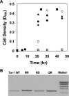
Figure 1
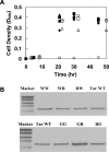
Figure 1. Membrane insertion and chimaeric protein expression
(A) Correct integration of the ToxR-TM–MalE chimaeric proteins was examined by their ability to functionally complement the MalE deficiency of PD28 cells. PD28 cells were transformed with the different plasmids and were grown in minimal medium containing maltose. Only cells that expressed periplasmic MalE were able to grow with maltose as the only carbon source. Tar-1 Q/R and R/S constructs showed similar growth curves as Tar-1 WT, indicating proper membrane integration. The double arginine mutation and the negative control with the deleted TM domain (ΔTM) showed no growth. ◆, Tar-1 WT; ○, GPA; ■, A16; □, Tar R/R; ▲, Q/R; ●, R/S; and ◇, ΔTM. (B) Comparison of the ToxR-TM–MalE chimaeric proteins' expression levels (shown by the bands at ≈65 kDa). Samples of FHK12 cells containing different sequences of Tar-1 within the ToxR–MalE chimaeric protein were lysed in SDS sample buffer, separated on SDS/12%-PAGE and immunoblotted using anti-(maltose binding protein) antibody (New England Biolabs). The chimaeric protein mutants showed expression levels similar to that of the WT TM domain. Sizes of the markers from the bottom to the top are 60, 80, 100, 140 and 200 kDa.
Figure 2
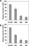
Figure 2. Arginine mutations of the Tar-1 WT TM domain
Cells expressing a ToxR-TM–MalE chimaera were examined for dimerization activity (normalized relative to the WT Tar-1 TM domain activity). All values are the average of at least three independent assays. Error bars represent the estimated S.D. The exact sequences are shown in Table 1.
Figure 3
Figure 3. Arginine mutations of the Tar-1 W/W and G/G TM domains
Cells expressing a ToxR-TM–MalE chimaera were examined for dimerization activity. The details are as described in the legend to Figure 2.
Figure 4
Figure 4. Membrane insertion and chimaeric protein expression of arginine mutants
(A) Correct integration of the ToxR-TM–MalE chimaeric proteins was examined as described in the legend to Figure 1(A). All constructs showed similar growth curves, except the negative control with the deleted TM domain (ΔTM), indicating proper membrane integration. ◆, Tar-1 WT; ▲, G/G; ●, R/G; ■, G/R; ○, W/W; △, R/W; □, W/R; and ◇, ΔTM. (B) Comparison of the ToxR-TM–MalE chimaeric proteins' expression levels (65 kDa). The details are as described in the legend to Figure 1(B).
Figure 5
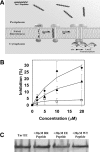
Figure 5. Tar-1 hetero-oligomerization
(A) Schematic illustration of the ToxR hetero-oligomerization system. The association of the Tar-1 TM domains activates ToxR, which only then can bind the ctx promoter and initiate the lacZ transcription process. Hetero-association of the exogenous peptides with the ToxR-Tar-1 TM domain prevents the activation of ToxR by shifting the equilibrium towards monomeric ToxR, thus reducing lacZ transcription and hence its signal. (B) Inhibition of the dimerization level in the presence of the Tar-1 WT, Tar-1 E/E and Tar-1 R/R peptides. The dose–response of β-gal inhibition as a function of exogenous peptide concentration: ▲, Tar-1 WT; ◆, Tar-1 R/R; and □, Tar-1 E/E. The results were normalized relative to the WT Tar-1 TM domain activity. All values are the average of at least three independent assays. Error bars represent the estimated S.D. (C) Comparison of the Tar-1 E/E chimaera expression levels in the presence of 20 μM of each peptide. The details are as described in the legend to Figure 1. The peptides had no significant effect on the expression of the Tar-1 E/E chimaeric protein.
Figure 6
Figure 6. CD spectra of the peptides in SDS micelles
Far-UV CD spectra of W/T (●), E/E (△) and R/R (■) Tar-1 TM domain peptides in 1% SDS. Spectra were measured on an Aviv spectropolarimeter at 0.2 nm intervals with a 10 s average time, using a 0.1 cm light path.
Figure 7
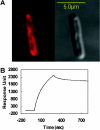
Figure 7. Localization of the Tar-1 peptides to the bacterial membrane
(A) Partitioning of rhodamine-labelled Tar-1 R/R to the inner membrane of E. coli was determined by confocal laser-scanning microscopy. A similar result was observed for the Tar-1 WT and Tar-1 E/E peptides. The Tar-1 WT peptide was previously shown to be localized to the bacterial membrane. The fluorescence image is on the upper-left-hand side and the transmission light image is on the upper-right-hand side. Most of the peptide is localized on the plasma membrane, although some peptide also penetrates into the cytoplasm. Confocal images were obtained using an Olympus IX70 FV500 confocal laser-scanning microscope. There were only minute background fluorescence intensities observed in the control bacteria. (B) A sensogram of the binding between Tar-1 WT peptide (10 μM) and PE/PG (7:3, w/w) bilayers.

Similar articles

Cited by

References

    1. Ding P. Z., Wilson T. H. The effect of modifications of the charged residues in the transmembrane helices on the transport activity of the melibiose carrier of Escherichia coli. Biochem. Biophys. Res. Commun. 2001;285:348–354. - PubMed
    1. Burnay M., Crambert G., Kharoubi-Hess S., Geering K., Horisberger J. D. Electrogenicity of Na,K- and H,K-ATPase activity and presence of a positively charged amino acid in the fifth transmembrane segment. J. Biol. Chem. 2003;278:19237–19244. - PubMed
    1. Xu Y., Kakhniashvili D. A., Gremse D. A., Wood D. O., Mayor J. A., Walters D. E., Kaplan R. S. The yeast mitochondrial citrate transport protein. Probing the roles of cysteines, Arg181, and Arg189 in transporter function. J. Biol. Chem. 2000;275:7117–7124. - PubMed
    1. Partridge A. W., Melnyk R. A., Deber C. M. Polar residues in membrane domains of proteins: molecular basis for helix-helix association in a mutant CFTR transmembrane segment. Biochemistry. 2002;41:3647–3653. - PubMed
    1. Partridge A. W., Therien A. G., Deber C. M. Missense mutations in transmembrane domains of proteins: phenotypic propensity of polar residues for human disease. Proteins. 2004;54:648–656. - PubMed

Publication types

MeSH terms