. 2004 Sep;165(3):719-28.
doi: 10.1016/s0002-9440(10)63335-4.

Glutathione depletion impairs myogenic differentiation of murine skeletal muscle C2C12 cells through sustained NF-kappaB activation

Affiliations

Glutathione depletion impairs myogenic differentiation of murine skeletal muscle C2C12 cells through sustained NF-kappaB activation

Esther Ardite et al. Am J Pathol. 2004 Sep.

Abstract

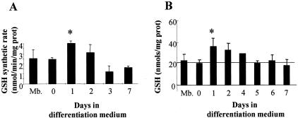
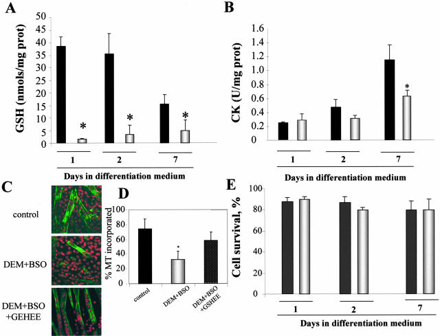
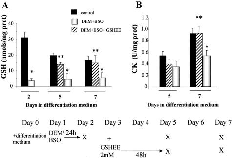
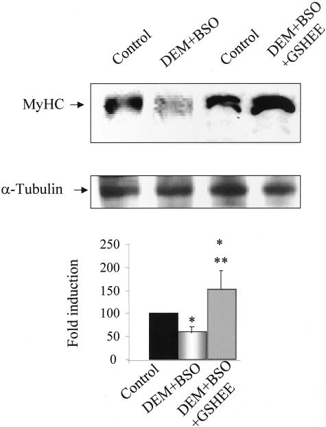
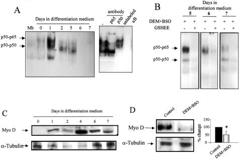
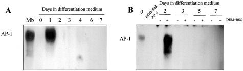
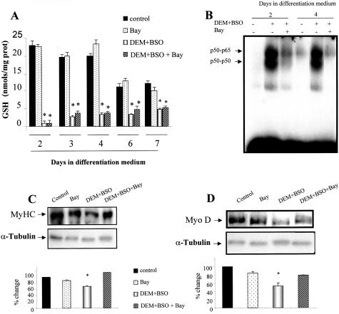
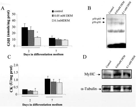
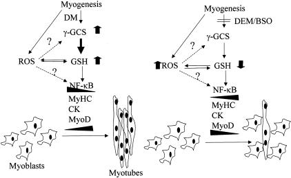
Skeletal muscle differentation is a complex process regulated at multiple levels. This study addressed the effect of glutathione (GSH) depletion on the transition of murine skeletal muscle C2C12 myoblasts into myocytes induced by growth factor inactivation. Cellular GSH levels increased within 24 hours on myogenic stimulation of myoblasts due to enhanced GSH synthetic rate accounted for by stimulated glutamate-L-cysteine ligase (also known as gamma-glutamylcysteine synthetase) activity. In contrast, the synthesis rate of GSH using gamma-glutamylcysteine and glutamate as precursors, which reflects the activity of the GSH synthetase, did not change during differentiation. The stimulation of GSH stores preceded the myogenic differentiation of C2C12 myoblasts monitored by expression of muscle-specific genes, creatine kinase (CK), myosin heavy chain (MyHC), and MyoD. The pattern of DNA binding activity of NF-kappaB and AP-1 in differentiating cells was similar both displaying an activation peak at 24 hours after myogenic stimulation. Depletion of cellular GSH levels 24 hours after stimulation of differentiation abrogated myogenesis as reflected by lower CK activity, MyHC levels, MyoD expression, and myotubes formation, effects that were reversible on GSH replenishment by GSH ethyl ester (GHSEE). Moreover, GSH depletion led to sustained activation of NF-kappaB, while GSHEE prevented it. Furthermore, inhibition of NF-kappaB activation restored myogenesis despite GSH depletion. Thus, GSH contributes to the formation of myotubes from satellite myoblasts by ensuring inactivation of NF-kappaB, and hence maintaining optimal GSH levels may be beneficial in restoring muscle mass in chronic inflammatory disorders.


Figures

Figure 1
Figure 1
Enhanced GSH synthetic rate during myogenesis. Myoblasts were grown to 100% confluency for 24 hours in growth medium and then the culture media switched to differentiation medium to induce differentiation. A: Cells were lysed and the cytosolic fraction was used to assess the maximal GSH synthetic rate in a cell-free system in the presence of unlimited GSH precursors (glutamate, cysteine, and glycine) and needed cofactors as described in Materials and Methods. The de novo synthesized GSH was conjugated with monochlorobimane and the fluorescence of the adduct was continuously determined over time in a fluorimeter. B: Total GSH content (GSH+GSSG) was determined in cell lysates from confluent myoblasts (Mb) and differentiating cells at different times of differentiation following culture in differentiation medium. Cell GSH content was analyzed by HPLC after derivatization with dinitrofluorobenzene as described in Materials and Methods. Data are the mean ± SD of three to six independent experiments. *, P < 0.05 versus Mb.
Figure 2
Figure 2
GSH depletion impairs differentiation of C2C12 cells. Myoblasts were grown in growth medium to reach confluency and 24 hours later the culture medium was switched to differentiation medium to induce myogenesis. Twenty-four hours after myogenic stimulation in differentiation medium cells were treated with DEM (0.8 mmol/L preincubation for 15 minutes) followed by BSO (1 mmol/L) for 24 hours to deplete GSH and inhibit γ-GCS activity (DEM+BSO, open bars) or not (control, closed bars), after which cells were grown in differentiation medium until day 7. A: Total GSH levels were assessed by HPLC as in Figure 1B. B: To assess the effect of DEM/BSO on differentiation and myogenesis, CK activity was determined in cell lysates at different times of culture in differentiation medium with or without DEM+BSO preincubation. C: The morphological appearance of myotubes at day 7 was examined by confocal microscopy. Nuclei were stained by propidium iodide (red) and MyHC by an anti-MyHC antibody followed by a FITC-labeled secondary antibody (green). In some cases we examined the effect of GSH replenishment by GSHEE on DEM+BSO-treated cells. D: Quantitation of myotube formation from the representative images shown in C as the percentage of nuclei in MyHC-positive cells compared to total number of nuclei in the field. E: Cell survival was determined after treatment with DEM+BSO as the percentage of LDH released into the medium and viability expressed as percentage of LDH found in cells plus medium. Data are the mean ± SD of three to five independent experiments. *, P < 0.05 versus untreated control cells cultured in DM medium.
Figure 3
Figure 3
Treatment with GSH ethyl ester restores differentiation of GSH-depleted C2C12 cells. Confluent myoblasts were treated with differentiation medium to induce differentiation with or without DEM+BSO treatment at day 1 for 24 hours as indicated in the bottom scheme. In some cases, DEM+BSO-pretreated cells were then exposed to GSH ethyl ester (GSHEE, 2 mmol/L) for 48 hours and cultured in fresh differentiation medium until day 7 as indicated. The X in the bottom scheme represents the time at which cells were taken for determination of total GSH levels (A) or CK activity as a marker for biochemical differentiation (B). Results are the mean ± SD of three to four independent experiments. *, P < 0.05 control; **, P < 0.04 versus DEM+BSO.
Figure 4
Figure 4
Regulation of MyHC expression by GSH levels. Myoblasts were stimulated for differentiation into myotubes by switching culture medium to differentiation medium. In some cases, cells were treated with DEM+BSO with or without GSHEE (2 mmol/L) as indicated in the scheme of Figure 3. At day 7 of culture in differentiation medium, cells were lysed and levels of MyHC or α-tubulin as a loading control were determined by Western blot. The bottom panel represents the quantitive effect of DEM+BSO with or without GSHEE on MyHC levels. Results are the mean ± SD of three to four independent experiments with similar results. *, P < 0.05 versus control; **, P < 0.05 versus DEM+BSO.
Figure 5
Figure 5
Regulation of NF-κB activation and MyoD expression during myogenesis by GSH. A: NF-κB activation was examined by electrophoretic mobility shift assay in nuclear extracts prepared from myoblasts or at different days after myogenesis induction by differentiation medium. The arrows denote the presence of the p65/p50 heterodimer and p50/p50 homodimer using anti-p65 and anti-p50 antibodies shown in the supershift assay. In addition the specificity of the complexes was ascertained by competition with a molar excess of unlabeled oligonucleotide. B: In some cases, cells were treated with DEM+BSO at day 1 of differentiation medium stimulation for 24 hours and then cultured with or without GSHEE (2 mmol/L) for 48 hours, isolating nuclear extracts from days 5 to 7 to examine NF-κB activation in nuclear extracts. C: The levels of MyoD and α-tubulin were determined in C2C12 cells at different times after culture in differentiation medium. D: Effect of DEM+BSO pretreatment at day 1 for 24 hours on the levels of MyoD and α-tubulin at day 2 of culture in differentiation medium. Data shown are representative of three independent experiments with similar results observed.
Figure 6
Figure 6
AP-1 activation during myogenesis with or without GSH depletion. A: AP-1 activation was examined by electrophoretic mobility shift assay in nuclear extracts from C2C12 cells at different times during differentation. B: Cells were treated with DEM+BSO at day 1 of differentiation isolating nuclear extracts at different times during differentiation to examine AP-1 activation. The cellular GSH levels after DEM+BSO addition remained depleted by 88% with respect to control cells from day 1 to day 7. Data shown are representative of four independent experiments with similar results observed.
Figure 7
Figure 7
Inhibition of NF-κB activation restores myogenesis despite GSH depletion. Differentiating C2C12 cells were treated with DEM+BSO at day 1 with or without Bay 11–7085 (Bay) to prevent NF-κB activation. A: GSH levels at different times of differentiation were determined as indicated in Figure 1B. B: NF-κB activation was examined in nuclear extracts from cells treated with DEM+BSO with or without Bay which prevents NF-κB activation by inhibition of IκBα phosphorylation. The effect of Bay on differentiation of DEM+BSO-treated cells was examined by the expression of MyHC (C) and MyoD (D) in cell extracts taken at day 7 after culture in differentiation medium with respect to the α-tubulin levels used as loading control. The changes quantitated in the corresponding panels are the mean ± SD of three independent experiments. *, P < 0.05 versus controls.
Figure 8
Figure 8
Mild GSH depletion does not impair myogenesis. Differentiating C2C12 cells were treated with DEM at the doses shown at day 1 for 15 minutes and then BSO was added as indicated in Figure 3. A: GSH levels were examined in differentiating cells at day 2 and 7 as indicated in Figure 1B. B: NF-κB activation was examined in nuclear extracts from cells pretreated with DEM at the doses indicated. The effect of mild GSH depletion on differentiation was examined by CK activity (C) as well as expression of MyHC with respect to α-tubulin (D).
Figure 9
Figure 9
γ-GCS induction during myogenesis. Schematic diagram depicting our findings. Myogenesis differentiation induces γ-GCS resulting in enhanced GSH levels. The signal and mechanism leading to induction of γ-GCS is uncertain and may include overgeneration of ROS possibly from mitochondria (see text for details). Enhanced GSH levels ensures myogenesis perhaps through down-regulation of NF-κB and subsequent induction of muscle-specific genes. On the other hand, the abolishment of stimulated GSH levels may contribute to persistent activation of NF-κB, which in turn contribute to the down-regulation of MyoD.

Similar articles

Cited by

References

    1. Kotler DP, Tierney AR, Wang J, Pierson RN., Jr Magnitude of body-cell-mass depletion and the timing of death from wasting in AIDS. Am J Clin Nutr. 1989;50:444–447. - PubMed
    1. Argiles JM, Lopez-Soriano FJ. The role of cytokines in cancer cachexia. Med Res Rev. 1999;19:223–248. - PubMed
    1. Schols AM, Slangen J, Volovics L, Wouters EF. Weight loss is a reversible factor in the prognosis of chronic obstructive pulmonary disease. Am J Respir Crit Care Med. 1998;157:1791–1797. - PubMed
    1. Zhao SP, Zeng LH. Elevated plasma levels of tumor necrosis factor in chronic heart failure with cachexia. Int J Cardiol. 1997;58:257–261. - PubMed
    1. Di Francia M, Barbier D, Mege JL, Orehek J. Tumor necrosis factor-α levels and weight loss in chronic obstructive pulmonary disease. Am J Respir Crit Care Med. 1994;150:1453–1455. - PubMed

Publication types

MeSH terms