. 2004 Sep;165(3):741-51.
doi: 10.1016/s0002-9440(10)63337-8.

Solar ultraviolet irradiation reduces collagen in photoaged human skin by blocking transforming growth factor-beta type II receptor/Smad signaling

Affiliations

Solar ultraviolet irradiation reduces collagen in photoaged human skin by blocking transforming growth factor-beta type II receptor/Smad signaling

Taihao Quan et al. Am J Pathol. 2004 Sep.

Abstract

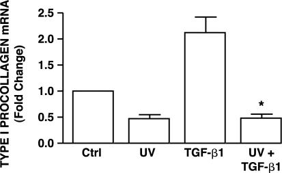
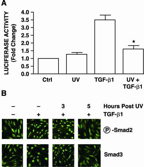
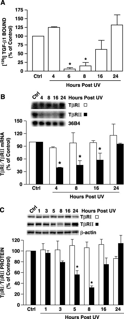
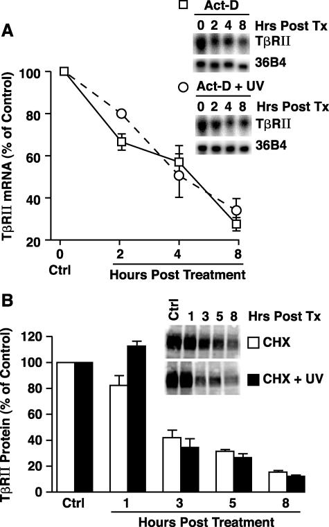
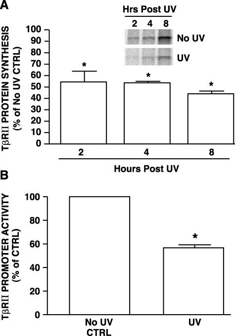
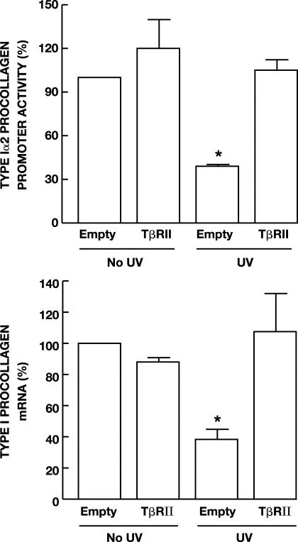
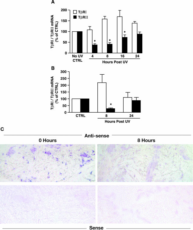
Ultraviolet (UV) irradiation from the sun reduces production of type I procollagen (COLI), the major structural protein in human skin. This reduction is a key feature of the pathophysiology of premature skin aging (photoaging). Photoaging is the most common form of skin damage and is associated with skin carcinoma. TGF-beta/Smad pathway is the major regulator of type I procollagen synthesis in human skin. We have previously reported that UV irradiation impairs transforming growth factor-beta (TGF-beta)/Smad signaling in mink lung epithelial cells. We have investigated the mechanism of UV irradiation impairment of the TGF-beta/Smad pathway and the impact of this impairment on type I procollagen production in human skin fibroblasts, the major collagen-producing cells in skin. We report here that UV irradiation impairs TGF-beta/Smad pathway in human skin by down-regulation of TGF-beta type II receptor (TbetaRII). This loss of TbetaRII occurs within 8 hours after UV irradiation and precedes down-regulation of type I procollagen expression in human skin in vivo. In human skin fibroblasts, UV-induced TbetaRII down-regulation is mediated by transcriptional repression and results in 90% reduction of specific, cell-surface binding of TGF-beta. This loss of TbetaRII prevents downstream activation of Smad2/3 by TGF-beta, thereby reducing expression of type I procollagen. Preventing loss of TbetaRII by overexpression protects against UV inhibition of type I procollagen gene expression in human skin fibroblasts. UV-induced down-regulation of TbetaRII, with attendant reduction of type I procollagen production, is a critical molecular mechanism in the pathophysiology of photoaging.


Figures

Figure 1
Figure 1
UV irradiation inhibits TGF-β1-induced type I procollagen gene expression in cultured human skin fibroblasts. Cells were sham or UV irradiated (30 mJ/cm2). Vehicle or TGF-β1 (5 ng/ml) was added 8 hours after irradiation, and cells were harvested 16 hours later. Type I procollagen mRNA levels were determined by real-time RT-PCR and were normalized to mRNA levels of the housekeeping gene 36B4. Data are presented as fold change in type I procollagen levels relative to nontreated control (Ctrl) cells, and are expressed as mean ± SEM, n = 3. *, P < 0.05, compared with TGF-β1 treatment.
Figure 2
Figure 2
UV irradiation inhibits TGF-β/Smad signaling pathway in cultured human skin fibroblasts. A: UV irradiation inhibits TGF-β/Smad-regulated reporter gene in human skin fibroblasts. Cells were co-transiently transfected with TGF-β/Smad-regulated luciferase reporter constructs (SBEX4) and β-galactosidase expression vector (used as internal control). Twenty-four hours after transfection, cells were sham or UV irradiated (30 mJ/cm2), and then 8 hours later, cells were treated with vehicle or TGF-β1 (5 ng/ml) for 16 hours. Aliquots containing identical β-galactosidase activity were used for each luciferase assay. Data are presented as fold change in luciferase activity relative to activity in nontreated control (Ctrl) cells and are expressed as mean ± SEM, n = 3. *, P < 0.05, compared with TGF-β1. B: UV irradiation inhibits TGF-β-induced Smad2 and Smad3 nuclear translocation in human skin fibroblasts. Cells were sham (−) or UV irradiated (30 mJ/cm2), and then treated at the indicated times after UV irradiation with vehicle (−) or TGF-β1 (+, 5 ng/ml) for 1 hour. Phosphorylated Smad2 (P-Smad2) and Smad3 nuclear translocation were determined by immunofluorescence confocal microscopy as described in Materials and Methods. Results are representative of three experiments.
Figure 3
Figure 3
UV irradiation reduces TβRII mRNA and protein in cultured human skin fibroblasts. A: UV irradiation inhibits TGF-β1 receptor binding in human skin fibroblasts. Cells were sham or UV irradiated (30 mJ/cm2). At the indicated times after UV specific binding of [125I]TGF-β1 to intact cells was determined as described in Materials and Methods. Data are presented as percentage of [125I]TGF-β1 binding relative to nonirradiated control (Ctrl) cells and are expressed as mean ± SEM, n = 4. *, P < 0.05, compared with control. B: UV irradiation reduces TβRII mRNA levels in human skin fibroblasts. Cells were sham or UV irradiated (30 mJ/cm2), total RNA was prepared at the indicated times after UV, and TβRI, TβRII, and 36B4 mRNA levels were determined by Northern analysis. Inset shows representative Northern blot. Data are presented as percentage of TβRI and TβRII levels relative to nonirradiated control (Ctrl) cells and are expressed as mean ± SEM, n = 3. *, P < 0.05, compared with control. C: UV irradiation reduces TβRII protein levels in human skin fibroblasts. Cells were sham or UV irradiated (30 mJ/cm2), membrane fractions were prepared at the indicated time after UV, and TβRI and TβRII protein levels in membrane fractions were quantified by Western analysis. Inset shows representative Western blot for TβRI, TβRII, and β-actin control. Data are presented as percentage of TβRI and TβRII levels relative to nonirradiated control cells (Ctrl) and are expressed as mean ± SEM, n = 3. *, P < 0.05, compared with control.
Figure 4
Figure 4
UV irradiation does not alter TβRII mRNA or protein stability. A: Fibroblasts were incubated with transcription inhibitor actinomycin D (Act-D, 10 μg/ml), either alone or in combination with UV irradiation (30mJ/cm2). Total RNA was isolated at the indicated times and subjected to Northern analysis. Data are presented as percentage of TβRII mRNA levels relative to vehicle-treated control cells (Ctrl), and are expressed as means + SEM, n = 3. B: Cells were incubated with translation inhibitor cycloheximide (CHX, 10 μg/ml), either alone or in combination with UV irradiation (30mJ/cm2). Whole cell extract was isolated at the indicated times and subjected to Western analysis. Data are presented as percentage of TβRII protein levels relative to vehicle-treated control cells (Ctrl) and are expressed as mean + SEM, n = 3.
Figure 5
Figure 5
UV irradiation inhibits TβRII protein synthesis and promoter transcription in human skin fibroblasts. A: UV irradiation inhibits TβRII protein synthesis in human skin fibroblasts. Cells were sham or UV irradiated and then immediately incubated with [35S]methionine/cysteine for the indicated times. [35S]-Labeled TβRII protein was immunoprecipitated and subjected to 12% sodium dodecyl sulfate-polyacrylamide gel electrophoresis. The bands represent [35S]-labeled TβRII protein synthesized during the indicated times. Bars are means ± SEM band intensities of TβRII protein levels in UV-irradiated cells, as a percentage in sham-irradiated cells. TβRII protein levels in sham-irradiated cells were assigned a value of 100% at each time point, n = 3. *, P < 0.05, compared with control. B: UV irradiation inhibits transcription of the TβRII promoter reporter in human skin fibroblasts. TβRII promoter/luciferase reporter (−1640/+62) was transiently co-transfected with β-galactosidase expression vector into the human skin fibroblasts. Twenty-four hours after transfection, cells were sham or UV irradiated (30 mJ/cm2). TβRII promoter activity was determined 8 hours after irradiation by luciferase assay. Aliquots containing identical β-galactosidase activity were used for each luciferase assay. Data are presented as percentage of control of promoter activity relative to activity in nonirradiated control cells (Ctrl) and are expressed as mean ± SEM, n = 3. *, P < 0.05, compared with control.
Figure 6
Figure 6
Overexpression of TβRII prevents UV inhibition of type I procollagen gene expression in human skin fibroblasts. A: Cells were transiently co-transfected with type I(α2) procollagen promoter CAT-reporter construct, β-galactosidase expression vector, and where indicated, empty or TβRII expression vector. Twenty-four hours after transfection, cells were sham or UV irradiated (30 mJ/cm2) and then 8 hours later treated with TGF-β1 (5 ng/ml) for 16 hours. Aliquots containing identical β-galactosidase activity were used for each procollagen promoter reporter activity assay. Data are presented as percentage of control in reporter CAT activity relative to empty vector control cells, and are expressed as mean ± SEM, n = 3. *, P < 0.05, compared to control. B: Human fibroblasts were transiently transfected with TβRII expression vector or empty vector (pCDNA 3.1). Forty-eight hours after transfection, cells were sham or UV irradiated (30 mJ/cm2). Sixteen hours after UV irradiation, type I(α1) procollagen and 36B4 (internal control) mRNA levels were quantified by real-time RT-PCR. Data are presented as percentage of type I(α1) procollagen mRNA levels (normalized to 36B4 mRNA levels) relative to nonirradiated empty vector control cells and are expressed as mean ± SEM, n = 4. *, P < 0.05, compared to control.
Figure 7
Figure 7
Solar-simulated UV irradiation reduces TβRII mRNA expression in human skin in vivo. A: Solar-simulated UV irradiation reduces TβRII mRNA in skin connective tissue. Sun-protected human skin was exposed to solar-simulated UV (2 minimum erythema dose). At the indicated times, full-thickness human skin samples were obtained and snap-frozen. Epidermis was removed from dermis by dissection with a scalpel, under a dissecting microscope. Total RNA was prepared from dermis. TβRI (open bars) and TβRII (filled bars) mRNA levels were quantified by real-time RT-PCR and were normalized to mRNA levels of the housekeeping gene 36B4. Data are presented as percentage of TβRI and TβRII levels relative to nonirradiated control skin and are expressed as mean ± SEM, n = 6. *, P < 0.05, compared to control. B: Solar-simulated UV irradiation reduces TβRII mRNA in human skin fibroblasts in vivo. Fibroblasts in nonirradiated and solar-simulated UV-irradiated skin were captured by LCM as described in Materials and Methods. TβRI (open bars) and TβRII (filled bars) mRNA levels were quantified by real-time RT-PCR and normalized to mRNA levels of the housekeeping gene 36B4. Data are presented as percentage of TβRI and TβRII mRNA levels relative to nonirradiated control skin (Ctrl), and are expressed as mean ± SEM, n = 3 to 4. *, P < 0.05, compared to control. C: Dermal cellular localization of TβRII mRNA after solar-simulated UV irradiation in human skin in vivo. Skin samples were obtained at the indicated times after solar-simulated UV irradiation, and TβRII mRNA expression was determined by anti-sense riboprobe in situ hybridization. Sense riboprobe yielded minimal background signal. Results are representative of six patients.

Similar articles

Cited by

References

    1. Gilchrest BA, Yaar M. Ageing and photoageing of the skin: observations and the cellular and molecular level. Br J Dermatol. 1992;127:25–30. - PubMed
    1. Kaminer MS. Photodamage: magnitude of the problem. Gilchrest BA, editor. Cambridge: Blackwell Science,; Photodamage. 1995:pp 1–11.
    1. Brash D, Rudolph J, Simon J, Lin A, McKenna G, Baden H, Halperin A, Ponten J. A role for sunlight in skin cancer: UV-induced p53 mutations in squamous cell carcinoma. Proc Natl Acad Sci USA. 1991;88:10124–10128. - PMC - PubMed
    1. Warren R, Gartstein V, Kligman A, Montagna W, Allendorf R, Ridder G. Age, sunlight, and facial skin: a histologic and quantitative study. J Am Acad Dermatol. 1991;25:751–760. - PubMed
    1. Fisher GJ, Wang ZQ, Datta SC, Varani J, Kang S, Voorhees JJ. Pathophysiology of premature skin aging induced by ultraviolet light. N Engl J Med. 1997;337:1419–1428. - PubMed

Publication types

MeSH terms