Vitamin D3 induces caspase-14 expression in psoriatic lesions and enhances caspase-14 processing in organotypic skin cultures
- PMID: 15331408
- PMCID: PMC1618612
- DOI: 10.1016/S0002-9440(10)63346-9
Vitamin D3 induces caspase-14 expression in psoriatic lesions and enhances caspase-14 processing in organotypic skin cultures
Abstract
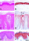
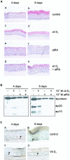
Caspase-14 is a nonapoptotic caspase family member whose expression in the epidermis is confined to the suprabasal layers, which consist of differentiating keratinocytes. Proteolytic activation of this caspase is observed in the later stages of epidermal differentiation. In psoriatic skin, a dramatic decrease in caspase-14 expression in the parakeratotic plugs was observed. Topical treatment of psoriatic lesions with a vitamin D3 analogue resulted in a decrease of the psoriatic phenotype and an increase in caspase-14 expression in the parakeratotic plugs. To investigate whether vitamin D3 directly affects caspase-14 expression levels, we used keratinocyte cell cultures. 1alpha,25-Dihydroxycholecalciferol, the biologically active form of vitamin D3, increased caspase-14 expression, whereas retinoic acid inhibited it. Moreover, retinoic acid repressed the vitamin D3-induced caspase-14 expression level. In addition, the use of organotypic skin cultures demonstrated that 1alpha,25-dihydroxycholecalciferol enhanced epidermal differentiation and caspase-14 activation, whereas retinoic acid completely blocked caspase-14 processing. Our data indicate that caspase-14 plays an important role in terminal epidermal differentiation, and its absence may contribute to the psoriatic phenotype.
Figures


Similar articles
-
Epidermal differentiation does not involve the pro-apoptotic executioner caspases, but is associated with caspase-14 induction and processing.Cell Death Differ. 2000 Dec;7(12):1218-24. doi: 10.1038/sj.cdd.4400785. Cell Death Differ. 2000. PMID: 11175259
-
Green tea polyphenol induces caspase 14 in epidermal keratinocytes via MAPK pathways and reduces psoriasiform lesions in the flaky skin mouse model.Exp Dermatol. 2007 Aug;16(8):678-84. doi: 10.1111/j.1600-0625.2007.00585.x. Exp Dermatol. 2007. PMID: 17620095
-
Caspase-14 expression by epidermal keratinocytes is regulated by retinoids in a differentiation-associated manner.J Invest Dermatol. 2002 Nov;119(5):1150-5. doi: 10.1046/j.1523-1747.2002.19532.x. J Invest Dermatol. 2002. PMID: 12445205
-
Caspase-14 reveals its secrets.J Cell Biol. 2008 Feb 11;180(3):451-8. doi: 10.1083/jcb.200709098. Epub 2008 Feb 4. J Cell Biol. 2008. PMID: 18250198 Free PMC article. Review.
-
Biological activity of vitamin D analogues in the skin, with special reference to antipsoriatic mechanisms.Br J Dermatol. 1995 May;132(5):675-82. doi: 10.1111/j.1365-2133.1995.tb00710.x. Br J Dermatol. 1995. PMID: 7772470 Review.
Cited by
-
Delphinidin, a dietary antioxidant, induces human epidermal keratinocyte differentiation but not apoptosis: studies in submerged and three-dimensional epidermal equivalent models.Exp Dermatol. 2013 May;22(5):342-8. doi: 10.1111/exd.12140. Exp Dermatol. 2013. PMID: 23614741 Free PMC article.
-
Luteolin induces caspase-14-mediated terminal differentiation in human epidermal keratinocytes.In Vitro Cell Dev Biol Anim. 2015 Nov;51(10):1072-6. doi: 10.1007/s11626-015-9936-5. Epub 2015 Oct 1. In Vitro Cell Dev Biol Anim. 2015. PMID: 26427706
-
Prodifferentiation, anti-inflammatory and antiproliferative effects of delphinidin, a dietary anthocyanidin, in a full-thickness three-dimensional reconstituted human skin model of psoriasis.Skin Pharmacol Physiol. 2015;28(4):177-88. doi: 10.1159/000368445. Epub 2015 Jan 22. Skin Pharmacol Physiol. 2015. PMID: 25620035 Free PMC article.
-
Tissue-specific and functional loci analysis of CASP14 gene in the sheep horn.PLoS One. 2024 Nov 21;19(11):e0310464. doi: 10.1371/journal.pone.0310464. eCollection 2024. PLoS One. 2024. PMID: 39570959 Free PMC article.
-
Lipid metabolism, apoptosis and cancer therapy.Int J Mol Sci. 2015 Jan 2;16(1):924-49. doi: 10.3390/ijms16010924. Int J Mol Sci. 2015. PMID: 25561239 Free PMC article. Review.
References
-
- Lavker RM, Sun TT. Epidermal stem cells. J Invest Dermatol. 1983;81:121s–127s. - PubMed
-
- Watt FM. Terminal differentiation of epidermal keratinocytes. Curr Opin Cell Biol. 1989;1:1107–1115. - PubMed
-
- Eckert RL, Crish JF, Robinson NA. The epidermal keratinocyte as a model for the study of gene regulation and cell differentiation. Physiol Rev. 1997;77:397–424. - PubMed
Publication types
MeSH terms
Substances
LinkOut - more resources
Full Text Sources
Other Literature Sources
Medical

