Comparative Study
. 2004 Sep;165(3):855-67.
doi: 10.1016/S0002-9440(10)63348-2.

WISP-1 is an osteoblastic regulator expressed during skeletal development and fracture repair

Affiliations
Comparative Study

WISP-1 is an osteoblastic regulator expressed during skeletal development and fracture repair

Dorothy M French et al. Am J Pathol. 2004 Sep.

Abstract

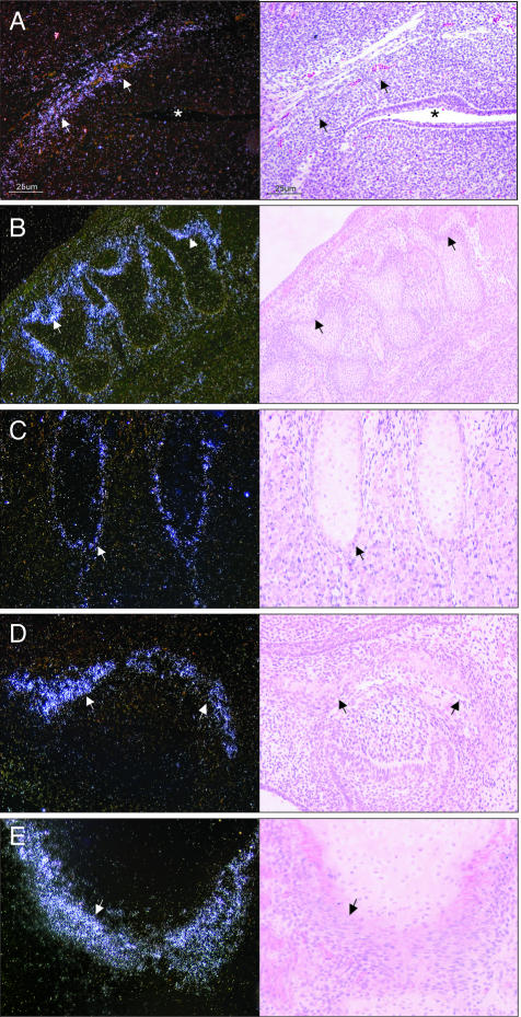
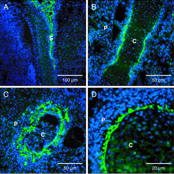
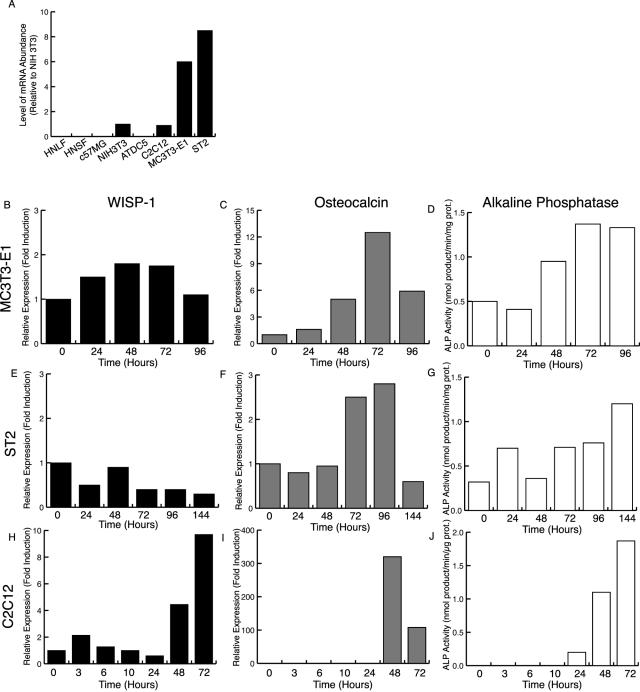
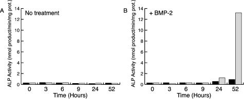
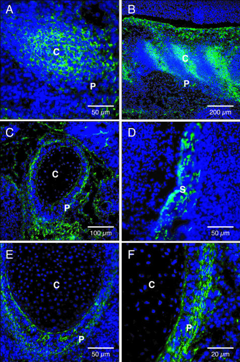
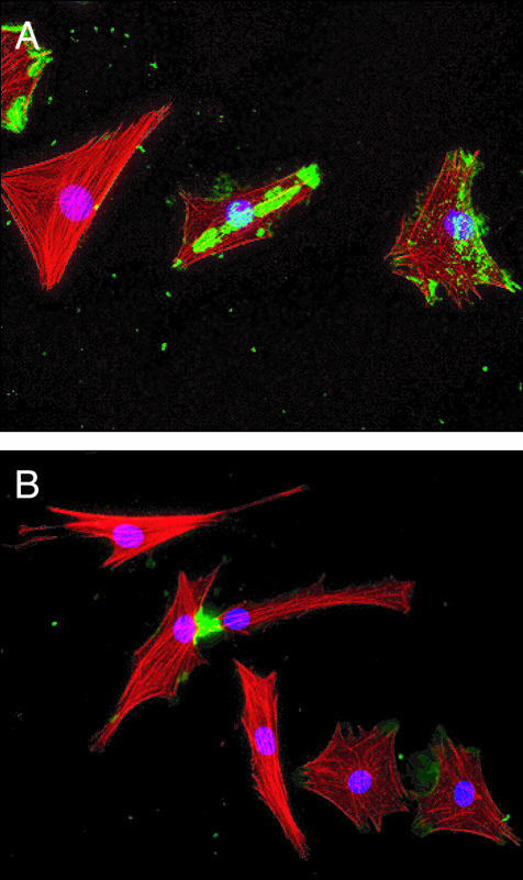
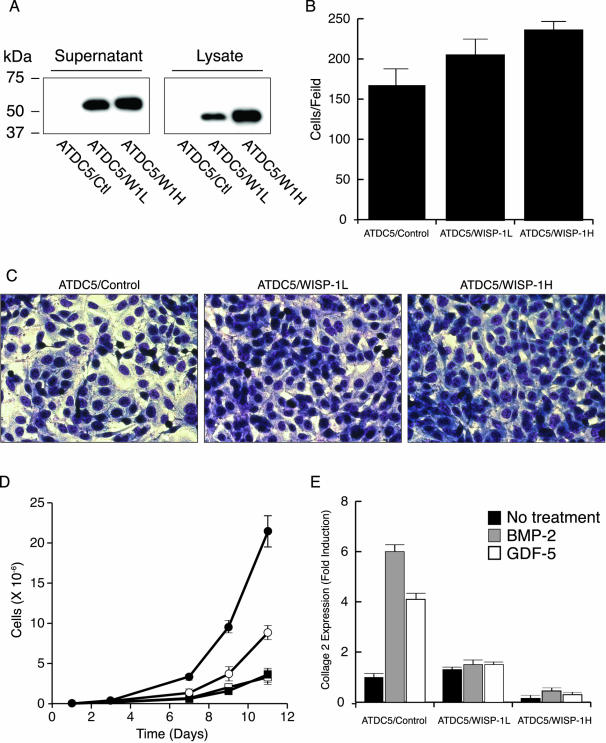
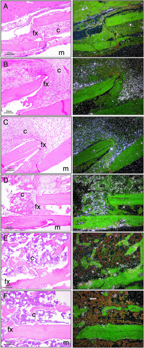
Wnt-1-induced secreted protein 1 (WISP-1) is a member of the CCN (connective tissue growth factor, Cyr61, NOV) family of growth factors. Experimental evidence suggests that CCN family members are involved in skeletogenesis and bone healing. To investigate the role of WISP-1 in osteogenic processes, we characterized its tissue and cellular expression and evaluated its activity in osteoblastic and chondrocytic cell culture models. During embryonic development, WISP-1 expression was restricted to osteoblasts and to osteoblastic progenitor cells of the perichondral mesenchyme. In vitro, we showed that WISP-1 expression in differentiating osteoblasts promotes BMP-2-induced osteoblastic differentiation. Using in situ and cell binding analysis, we demonstrated WISP-1 interaction with perichondral mesenchyme and undifferentiated chondrocytes. We evaluated the effect of WISP-1 on chondrocytes by generating stably transfected mouse chondrocytic cell lines. In these cells, WISP-1 increased proliferation and saturation density but repressed chondrocytic differentiation. Because of the similarity between skeletogenesis and bone healing, we also analyzed WISP-1 spatiotemporal expression in a fracture repair model. We found that WISP-1 expression recapitulates the pattern observed during skeletal development. Our data demonstrate that WISP-1 is an osteogenic potentiating factor promoting mesenchymal cell proliferation and osteoblastic differentiation while repressing chondrocytic differentiation. Therefore, we propose that WISP-1 plays an important regulatory role during bone development and fracture repair.


Figures

Figure 1
Figure 1
In situ hybridization of WISP-1 expression during mouse development. Left: Dark-field images; right: corresponding bright-field images. A: Base of the skull dorsal of the oropharynx (*) at E12.5. At E15.5, WISP-1 is expressed in osteoblasts and mesenchymal cells adjacent to bones undergoing endochondral ossification (B, vertebras; C, ribs) and intramembranous ossification (D, ossification within palatal shelf of maxilla). WISP-1 expression was similarly distributed in human embryo lower limb (E, lateral border of head of tibia). Original magnifications: ×100 (A, D); ×40 (B); ×200 (C, E).
Figure 2
Figure 2
Immunofluorescent localization of WISP-1 in rat embryo E18. Differentiating osteoblasts lining the calvaria (A), femur (B), and ribs (C, D). S, skull; P, periosteum; C, cartilage primordium. Original magnifications: ×100 (A); ×200 (B, C); ×400 (D).
Figure 3
Figure 3
WISP-1 is induced in differentiating osteoblasts. A: WISP-1 expression in different cell types. WISP-1 (B, E, H) and osteocalcin expression (C, F, I) and alkaline phosphatase activity (D, G, J) in MC3T3-E1 cells after ascorbic acid treatment (B–D), in ST2 cells after BMP-2 treatment (E–G), and in C2C12 cells after BMP-2 treatment (H–J).
Figure 4
Figure 4
WISP-1 promotes BMP-2-induced osteoblastic differentiation. C2C12 cells were transiently transfected with an empty vector (black bars) or WISP-1 expression construct (gray bars). Forty-eight hours after transfection, the culture media was replaced by media containing 5% FBS (A) or media containing 5% FBS and 300 ng/ml of BMP-2 (B) and alkaline phosphatase activity was measured at the indicated time.
Figure 5
Figure 5
In situ WISP-1 binding in rat embryo. At E14, WISP-1 binding revealed an intense fluorescent signal associated with costal (A) and vertebral (B) condensed mesenchymal cells. At E18, WISP-1 bound to osteoblasts and perichondral mesenchyme of developing bones; mesenchyme surrounding cartilage primordium of rib (C), calvaria (D), mesenchyme surrounding cartilage primordium of distal part of radius (E, F). P, perichondrium; C, cartilage primordium; S, skull. Original magnifications: ×200 (A, D, E); ×40 (B); ×100 (C); ×400 (F).
Figure 6
Figure 6
WISP-1 binding to dedifferentiated chondrocytes. A: The binding of WISP-1 (green) to dedifferentiated primary porcine chondrocytes showed an irregular pattern associated with patches and point of focal adhesion. B: Intense staining was found at the point of contact of adjacent cells. Red, actin filament staining; blue, nuclear staining. Original magnifications, ×200.
Figure 7
Figure 7
WISP-1 represses chondrogenic differentiation of ATDC5 cells. A: Western blot of WISP-1 produced by the ATDC5/control, ATDC5/WISP-1L, and ATDC5/WISP-1H cell lines. Saturation density (B) and photomicrograph (C) of ATDC5 cell lines grown to confluency. D: Proliferation of ATDC5 (open squares), ATDC5/control (filled squares), ATDC5/WISP-1L (open circles), and ATDC5/WISP-1H cells (filled circles). E: Relative expression of collagen 2 in ATDC5/control, ATDC5/WISP-1L, and ATDC5/WISP-1H cells before (black bars) and after inducing chondrocytic differentiation by BMP-2 (gray bars) or GDF-5 (white bars).
Figure 8
Figure 8
In situ hybridization of WISP-1 expression during fracture repair. Left: Bright-field images; right: corresponding dark-field images. Photomicrographs showing the localization of WISP-1 expression at days 3 (A), 5 (B), 7 (C), 14 (D), 21 (E), and 28 (F) after fracture. Each image is oriented with the medullary cavity (m) at the bottom right; the cortex, fracture (fx), and callus (c) occupy the majority of the photomicrograph. Original magnifications, ×100.

Similar articles

Cited by

References

    1. Bork P. The modular architecture of a new family of growth regulators related to connective tissue growth factor. FEBS Lett. 1993;327:125–130. - PubMed
    1. Brigstock DR. The connective tissue growth factor/cysteine-rich 61/nephroblastoma overexpressed (CCN) family. Endocr Rev. 1999;20:189–206. - PubMed
    1. Pennica D, Swanson TA, Welsh JW, Roy MA, Lawrence DA, Lee J, Brush J, Taneyhill LA, Deuel B, Lew M, Watanabe C, Cohen RL, Melhem MF, Finley GG, Quirke P, Goddard AD, Hillan KJ, Gurney AL, Botstein D, Levine AJ. WISP genes are members of the connective tissue growth factor family that are up-regulated in wnt-1-transformed cells and aberrantly expressed in human colon tumors. Proc Natl Acad Sci USA. 1998;95:14717–14722. - PMC - PubMed
    1. Perbal B. NOV (nephroblastoma overexpressed) and the CCN family of genes: structural and functional issues. Mol Pathol. 2001;54:57–79. - PMC - PubMed
    1. Xu L, Corcoran RB, Welsh JW, Pennica D, Levine AJ. WISP-1 is a Wnt-1- and beta-catenin-responsive oncogene. Genes Dev. 2000;14:585–595. - PMC - PubMed

Publication types

MeSH terms