Comparative Study
. 2004 Sep;165(3):889-99.
doi: 10.1016/S0002-9440(10)63351-2.

Angiopoietin-1 causes reversible degradation of the portal microcirculation in mice: implications for treatment of liver disease

Affiliations
Comparative Study

Angiopoietin-1 causes reversible degradation of the portal microcirculation in mice: implications for treatment of liver disease

Nicole L Ward et al. Am J Pathol. 2004 Sep.

Abstract

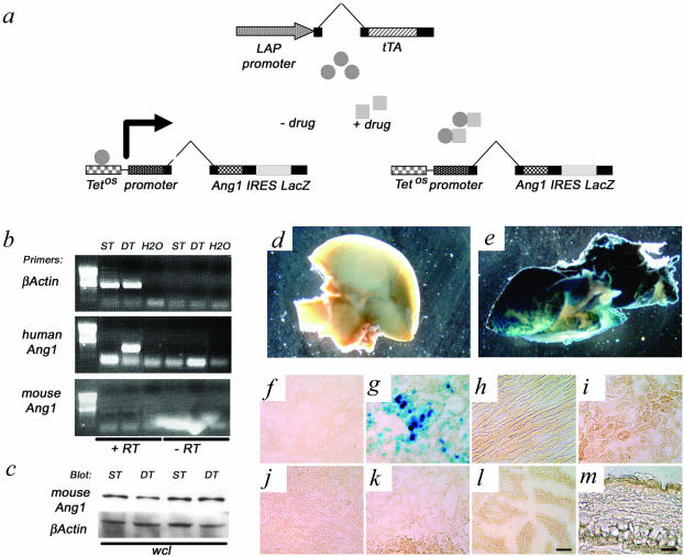
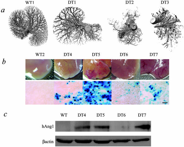
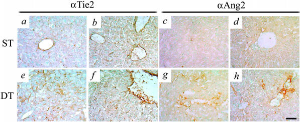
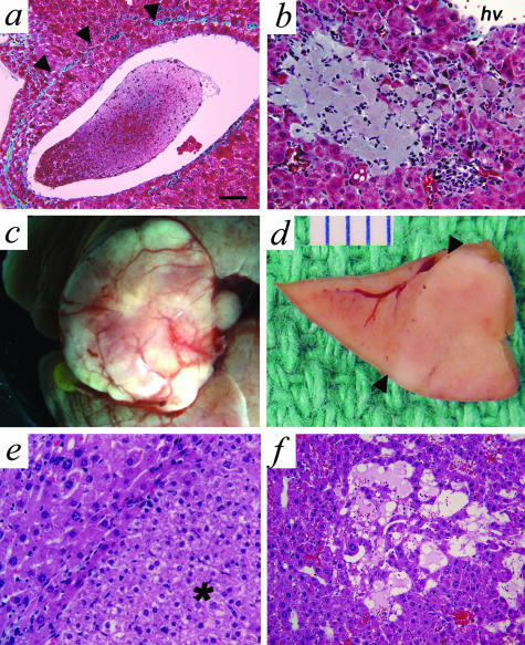
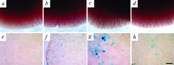
In many different liver diseases, such as cirrhosis, degradation of the microcirculation, including obliteration of small portal or hepatic veins contributes to disease-associated portal hypertension. The present study demonstrates the importance of angiogenesis in the establishment of arteriovenous shunts and the accompanying changes to the venous bed. One aspect of angiogenesis involves the branching of new vessels from pre-existing ones, and the molecular mechanisms controlling it are complex and involve a coordinated effort between specific endothelial growth factors and their receptors, including the angiopoietins. We modulated the hepatic vasculature in mice by conditionally expressing angiopoietin-1 in hepatocytes. In mice exposed to angiopoietin-1 during development, arterial sprouting, enlarged arteries, marked loss of portal vein radicles, hepatic vein dilation, and suggestion of arteriovenous shunting were observed. Most importantly, these phenotypic changes were completely reversed within 14 days of turning off transgene expression. Expression of excess angiopoietin-1 beginning in adulthood did not fully recapitulate the phenotype, but did result in enlarged vessels. Our findings suggest that controlling excessive angiogenesis during liver disease may promote the restoration of the portal vein circuit and aid in the resolution of disease-associated portal hypertension.


Figures

Figure 1
Figure 1
Generation and characterization of liver-specific Ang-overexpressing mice. a: Two independent lines of mice were used to induce the conditional expression of Ang-1 IRES LacZ. The tetracycline-responsive transactivator (tTA), expressed from the LAP promoter (driver transgene) associates with the tTA-binding site (TetOS) upstream of the human Ang-1 cDNA (responder transgene), which was bi-cis-tronically linked to the E. coli LacZ gene via an internal ribosomal entry site (IRES). In the absence of a tetracycline analog (−drug), Ang-1 is expressed and in the presence of this drug (+drug) Ang-1 expression in suppressed. b: RT-PCR expression analyses demonstrate the presence of human Ang-1 in the absence of Dox and that endogenous mouse Ang-1 levels do not dramatically change; +/− RT, presence or absence of reverse transcriptase, MMLV. c: Whole cell lysates were blotted with an antibody recognizing mouse Ang-1 and not human Ang-1, confirming that endogenous mouse Ang-1 protein levels do not change in the presence of the transgenic Ang-1. e and g: X-Gal staining in pieces and sections of double-transgenic (DT) adult liver showed strong expression of LacZ. d and f: No staining was observed in the single-transgenic (ST, d) or wild-type (WT, not shown) livers. LacZ expression in all DT mice was confined to the liver because no LacZ staining was observed in the heart (h), kidney (i), spleen (j), lung (k), intestine (l), or skin (m) of the ear. Scale bars: 100 μm (l); 50 μm (m).
Figure 2
Figure 2
DT mice with more severe phenotypes express more transgenic Ang-1 protein. a: Micro-CT images demonstrate overall vascular phenotypes of WT and DT livers. Some DT livers (DT1) appeared remarkably normal whereas others showed a range of severity (DT2 to DT3). WT and ST mouse (ST shown) livers at the gross morphological level (middle row, b) display the characteristic smooth capsular surface with no visible veins, consistent with normal liver morphology whereas the DT (DT4 to DT7) livers show a range of phenotype consisting of small to very large increases in dilatation of veins under the capsular surface. The range of phenotype corresponds to levels of LacZ staining (bottom row of b) and levels of transgenic Ang-1 protein expressed in the livers of these mice (c), such that mice with severe phenotypes (DT5 and DT7) have more LacZ staining and express more transgenic Ang-1 protein. Scale bar, 100 μm.
Figure 3
Figure 3
Ang-1 expression leads to alterations in hepatic vein architecture. ST and WT mouse livers (a) display the characteristic smooth capsular surface with no visible veins, consistent with normal liver morphology whereas DT livers (b) show a clear dilatation of veins under the capsular surface. Micro-CT images demonstrate even filling and the presence of portal and hepatic veins in WT and ST livers (c and e) and an obvious dilation of the hepatic veins and apparent reduction in the filling of the portal veins in DT livers (d and f). Interestingly, the dilated veins in the DT livers run parallel to the capsular surface (f) compared to the characteristic branching pattern displayed in the normal liver (e). At the microscopic level, WT and ST livers (g) stained with Masson Trichrome contain uniform portal and hepatic veins without any visible dilatation. h: DT livers present with a discernible dilatation of hepatic veins and an irregular distribution. a, c, e, and g are ST mice. Scale bar, 100 μm.
Figure 4
Figure 4
Ang-1-induced angiogenic events lead to liver reorganization. a: Masson Trichrome-stained sections from WT and ST livers demonstrate the characteristic pattern of organization of the portal tracts in proximity to a hepatic vein. b: All portal tracts in the WT liver contained a portal vein, portal artery, and a duct. c: In contrast, DT livers had disorganized portal tracts such that a marked enlargement of arteries was observed with very small portal veins or in smaller portal tracts, a complete absence of portal vein association. d: Higher magnification further demonstrates the absence of portal veins in two independent portal tracts. e–f: Arterial sprouts were observed without any associated portal veins, and these arterial twigs were localized closely with hepatic veins, suggesting the occurrence of AV shunts. a, artery; d, duct; hv, hepatic vein; pv, portal vein; pt, portal tract. Scale bars: ∼65 μm (a, c, e); ∼33 μm (b, d, and f).
Figure 5
Figure 5
Ang-1-induced increases in Tie2 and Ang-2 expression in ECs. ECs in ST and WT mice hepatic veins and portal veins and arteries express the Ang receptor Tie2 (a and b), and very low basal levels of Ang-2 (c and d). e and f: Tie2 levels increase in DT mice undergoing angiogenic remodeling, and are seen in hepatic veins and AV sprouts. g and h: Significant increases in Ang-2 levels are observed in DT mice and appear to occur in vessels undergoing angiogenic remodeling. Scale bar, 50 μm.
Figure 6
Figure 6
In severely affected DT mice, Ang-1-induced angiogenic events lead to hepatic vein remodeling, cell death, and nodular growths. a: Hepatic vein remodeling was seen as indicated by the presence of the original tunica media marked by a band of collagen (arrowheads). The tunica intima contains several layers of hepatocytes that encroach on the lumen. b: Small infarcts were observed in regions with absent portal veins. c: Some livers contained hyperplastic or neoplastic growths resembling FNH. d: In cross-section the tumor mass sits adjacent to normal-appearing liver tissue (arrowheads indicates the boundary) and is ∼1 cm in diameter. e: At the histological level, the margin of the tumor in e can be seen (arrowhead) and the tumor itself (*) is composed of normal-appearing hepatocytes without atypia. f: Within the region of the tumor a striking dilatation of the sinusoids, referred to as peliosis hepatis is seen. Scale bars: ∼65 μm (a); ∼33 μm (b, e, and f).
Figure 7
Figure 7
Ang-1-induced angiogenic events are reversible. a: Western blot analyses done on ST and DT mice before Dox exposure demonstrated the presence of transgenic Ang-1 in the DT mouse liver. After exposure to Dox (+Dox), transgenic Ang-1 production was suppressed. Three individual samples are shown (DT1 to DT3). b–d: Histological examination of DT mice having Ang-1 expression suppressed for 2 weeks with Dox returned the liver morphology back to normal with a uniform redistribution of veins. c and d: Histologically, normal portal tracts and normal hepatic veins without dilatation were observed in DT livers and the portal tracts contained veins, arteries, and ducts. e: Alternating regions of atrophic and nonatrophic hepatocytes were seen resembling the pattern of NRH, which may account for the mechanism of reversal. a, artery; d, duct; hv, hepatic vein; pv, portal vein; pt, portal tract. Scale bars: ∼65 μm (b and d); ∼33 μm (c).
Figure 8
Figure 8
Suppressing Ang-1 production during development and subsequent exposure to excess Ang-1 in adulthood leads to moderate levels of vascular remodeling. a and e: Mice exposed to Dox throughout gestation and into adulthood show no production of Ang-1 (as indicated by LacZ staining) and have completely normal looking livers. Transgenic Ang-1 production returns (as indicated by LacZ staining) 4 weeks (b, f), 8 weeks (c, g), and 14 weeks (d, h) after the removal of Dox exposure. Mild to moderate changes in the vasculature are observed at the edges of the liver as evidenced by moderate increases in vessel visibility and vessel size. No gross changes in vessel reorganization in the subcapsular space were seen in any of the DT livers.

Similar articles

Cited by

References

    1. Huet PM, Villeneuve JP, Pomier-Layrargues G, Marleau D. Hepatic circulation in cirrhosis. Clin Gastroenterol. 1985;14:155–168. - PubMed
    1. Wanless IR. Epithelioid hemangioendothelioma, multiple focal nodular hyperplasias, and cavernous hemangiomas of the liver. Arch Pathol Lab Med. 2000;124:1105–1107. - PubMed
    1. Wanless IR, Mawdsley C, Adams R. On the pathogenesis of focal nodular hyperplasia of the liver. Hepatology. 1985;5:1194–1200. - PubMed
    1. Wanless IR. Benign liver tumors. Clin Liver Dis. 2002;6:513–526. - PubMed
    1. Tanaka M, Wanless IR. Pathology of the liver in Budd-Chiari syndrome: portal vein thrombosis and the histogenesis of veno-centric cirrhosis, veno-portal cirrhosis, and large regenerative nodules. Hepatology. 1998;27:488–496. - PubMed

Publication types

MeSH terms

LinkOut - more resources