Comparative Study
. 2004 Sep;165(3):901-12.
doi: 10.1016/S0002-9440(10)63352-4.

Near completely humanized liver in mice shows human-type metabolic responses to drugs

Affiliations
Comparative Study

Near completely humanized liver in mice shows human-type metabolic responses to drugs

Chise Tateno et al. Am J Pathol. 2004 Sep.

Abstract

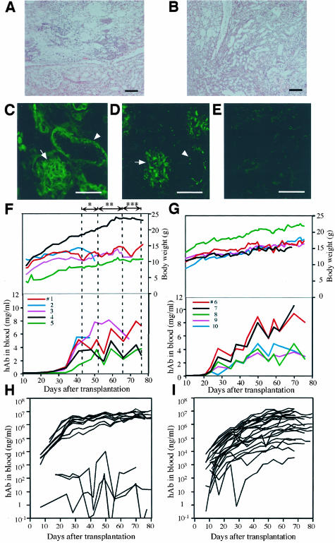
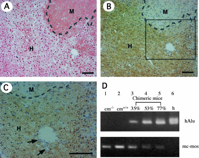
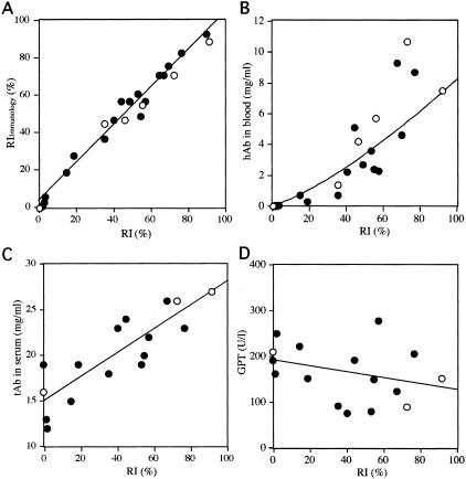
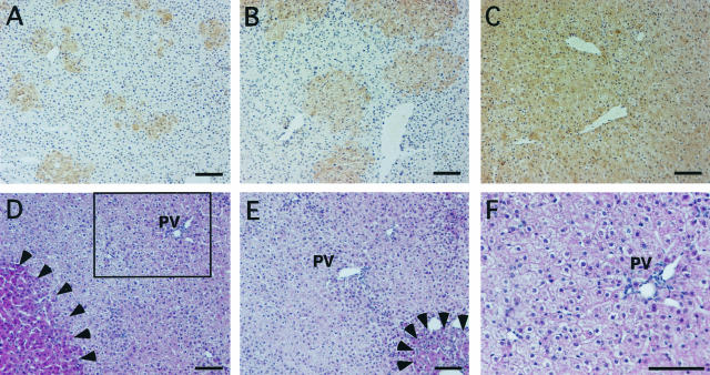
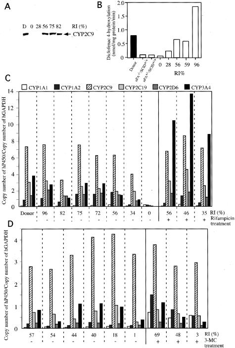
Human hepatocytes were transplanted into urokinase-type plasminogen activator-transgenic SCID mice (uPA/SCID mice), which are immunodeficient and undergo liver failure. The transplanted cells were characterized in terms of their in vivo growth potential and functions. The human hepatocytes progressively repopulated the murine host liver. However, the recipients died when the replacement index (RI) of the human hepatocytes exceeded 50%. The hosts (chimeric mice) survived at RI >50% when treated with a drug that has anti-human complement factor activity, and these mice developed livers with RI values as high as 96%. In total, 36 chimeric mice were generated, and the rate of successful engraftment was as high as 92%. The yield of chimeric mice with RI >70% was 32%. The human hepatocytes in the murine host liver expressed mRNAs for a variety of human cytochrome P450 (hCYP) subtypes, in a manner that was similar to the donor liver. The mRNAs for hCYP3A4 and hCYP1A1/2 were induced in the liver in a CYP type-specific manner when the mice were treated with rifampicin and 3-methylcholanthrene, respectively. These results indicate that human hepatocytes that propagate in mice retain their normal pharmacological responses. We conclude that the chimeric mouse developed in the present study is a useful model for assessing the functions and pharmacological responses of human hepatocytes.


Figures

Figure 1
Figure 1
Generation of humanized mice by treatment with or without a complement inhibitor. Histopathology (H&E staining) of the right kidney (A) and left kidney (B), and immunohistochemistry for hC3 (C) and hMAC (D), which was performed on the right kidney of a dying chimeric mouse that did not receive the complement inhibitor. The mouse that had a blood level of <3 mg/ml hAb and showed reduced body weight, was about to die 63 days after transplantation. Paraffin sections of the kidney were stained with H&E. A: Necrosis in renal papilla is evident in the kidney. B: Chronic pyelonephritis is observed throughout the kidney. hC3 (C) and hMAC (D) are deposited in the kidney. Photograph E represents the negative control stain for D. We confirmed that the antibodies against hC3 and hMAC did not cross-react with complement-induced necrotic tissues in the mouse ischemia/reperfusion kidney model. F: Relationships between body weights, hAb levels, and Futhan treatment. F-T hepatocytes of 12YM were transplanted into five mice (animals 1 to 5). The Futhan treatment schedule, which was determined by observing the changes in body weight of animal 1, was as follows: once every 2 days (*), once per day (**), and twice per day (***), as indicated by the double-arrowhead lines. The remaining four chimeric mice followed this regimen. Each line represents a chimeric mouse with the indicated animal number (#). G: Relationships between body weights, hAb levels, and Futhan regimen. The six mice in experiment 1 (Table 2) were transplanted with 12YM hepatocytes. Five of these mice were initially given Futhan on days when the hAb level was >2 mg/ml, and were administered thereafter with Futhan in the once every 2 days manner until the hAb level reached 4 mg/ml. Subsequently, Futhan was administered once per day or twice per day as long as the hAb concentration was between 4 and 6 mg/ml or >6 mg/ml, respectively. H: Humanized mice with 12 YM hepatocytes. The results obtained in experiments 1 and 2 are summarized in the graph. I: Humanized mice with 9MM hepatocytes. The uPA+/+/SCID+/+ mice were transplanted with 9MM hepatocytes (experiments 3 to 6 in Table 2). The mice were treated with Futhan according to the regimen described in G. Scale bars: 100 μm (A, B); 50 μm (C–E).
Figure 2
Figure 2
Demonstration of mouse liver chimerism. Histological serial sections were prepared from six liver lobes of each of 6 chimeric mice (donor, 12YM) and 19 chimeric mice (donor, 9MM), and stained with human genomic probes and anti-hCK8/18 antibodies. A: In situ hybridization of chimeric liver sections with human genomic DNA probes. Regions that contain hepatocytes with positive and negative nuclei are defined as human (H) and mouse (M) areas, respectively, the boundary being indicated by a dashed line. The RI of this chimeric mouse liver, which is calculated as the frequency of positive regions relative to that of the entire examined area in the sections, is 75%. B: CK8/18 immunostaining of a serial section of A. H and M indicate the regions that contain hepatocytes that are immunologically positive and negative, respectively, the boundary being indicated by a dashed line. The RI of this chimeric mouse liver, which is calculated as the frequency of immunopositive regions relative to that of the entire examined area in the sections, is given as RIImmunology = 77%, which is almost identical to the RI value calculated in A. C: High magnification of the region enclosed by the square in B. Bile duct cells, which are indicated with arrows, were negative for immunoreactivity. D: Chimerism determined by PCR. The hAlu sequence was amplified successfully from the liver genomic DNAs of human individuals (h, lane 6), and of chimeric mice with RI = 35% (lane 3), RI = 53% (lane 4), and RI = 77% (lane 5), but not from the control uPA−/−/SCID+/+ mouse (cm−/−; lane 1) or the control uPA+/+/SCID+/+ mouse (cm+/+; lane 2). PCR products for mc-mos were amplified from the mouse livers (lanes 1 and 2), but not from the human liver (lane 6). Scale bars, 100 μm.
Figure 3
Figure 3
Improvements in host liver function after repopulation with human hepatocytes. A: Correlation between the RIs, which were calculated using in situ hybridization and the RIImmunology values obtained by immunostaining with anti-CK8/18, for 6 animals (donor, 12YM; open circles) and 19 animals (donor, 9MM; filled circles). These animals are listed in Table 2. The two RI values are almost identical. The relationship between RIImmunology (y) and RI (x) fits the formula: y = 1.0 x + 3.2, with r2 = 0.98. B: The hAb concentration is plotted against the corresponding RI for the same animals as in A. C: The serum tAb concentrations are plotted against the corresponding RI values for three animals (12YM, open circles) and 13 animals (9MM, filled circles). D: The serum GPT levels are plotted against the corresponding RI values for the same animals as in C. The solid lines represent correlative curves.
Figure 4
Figure 4
Time course of repopulation of human hepatocytes in the host liver, and histopathology of the chimeric liver. Liver sections from mice that were transplanted with 9MM hepatocytes were stained with the anti-CK8/18 antibodies on days 14 (A), 35 (B), and 81 (C) after transplantation, at which time points the RIImmunology values were 10, 33, and 92%, respectively. The colonies that were derived from the human hepatocytes increased gradually in size, and were almost confluent by day 81 after transplantation. D to F: Histopathology of chimeric livers. H&E sections were prepared from the liver of a chimeric mouse with RI = 90% at 68 days after transplantation (donor 9MM). Most of the regions are populated with few eosinophilic human hepatocytes. There are a few nodules of transgene-deleted mouse hepatocytes, as indicated by the arrowheads in D. Some of the mouse hepatocytes in the degenerative regions that were basophilic originally have now become eosinophilic, as indicated by the arrowheads in E. The region enclosed by the square in D is magnified and shown in F. PV, portal vein. Scale bars, 100 μm.
Figure 5
Figure 5
Expression of hCYPs. A: Western blots for the detection of hCYP2C9. Western blotting was performed on the microsomes of the donor and 10 chimeric mice (donor, 12YM) using antibodies against hCYP2C9. The results are shown for the donor (D) and five chimeric mice with different RIs as examples. B: Measurements of liver diclofenac 4′-hydroxylation activities. Microsomes were isolated from the donor liver (filled bar), uPA−/−/SCID+/+, and uPA+/−/SCID+/+ control mouse livers (open bar), and five chimeric mouse livers (open bars with RI values), and were treated with diclofenac to measure their diclofenac 4′-hydroxylation activities. C and D: Profiles of the relative mRNA expression levels of six isoforms of hCYP. The ratios were obtained by dividing the copy number of each isoform mRNA in the donor and chimeric livers by that of hGAPDH mRNA in the donor and chimeric livers, respectively. The RI of each animal is indicated. C: Ten mice were transplanted with human hepatocytes (donor 12YM). Three mice (RI values of 56%, 46%, and 35%) were injected daily for 4 days with rifampicin, as indicated by +. The − designation represents mice that did not receive rifampicin treatment. D: Nine mice were transplanted with 9MM hepatocytes, three (RI values of 69%, 48%, and 3%) of which were injected daily for 4 days with 3-MC (indicated by +) before sacrifice for the measurement of CYP-specific mRNAs, whereas the other animals were not treated with 3-MC (indicated by −). The RI values indicated in A through D were calculated based on the percentage occupancy of in situ hybridization-positive regions in the histological sections of chimeric liver tissues.

References

    1. Dandri M, Burda MR, Török E, Pollok JM, Iwanska A, Sommer G, Rogiers X, Rogler CE, Gupta S, Will H, Greten H, Petersen J. Repopulation of mouse liver with human hepatocytes and in vivo infection with hepatitis B virus. Hepatology. 2001;33:981–988. - PubMed
    1. Mercer DF, Schiller DE, Elliott JF, Douglas DN, Hao C, Rinfret A, Addison WR, Fischer KP, Churchill TA, Lakey JRT, Tyrrell DLJ, Kneteman NM. Hepatitis C virus replication in mice with chimeric human livers. Nat Med. 2001;7:927–933. - PubMed
    1. Hamatani K, Matsuda Y, Araki R, Itoh M, Abe M. Cloning and chromosomal mapping of the mouse DNA-dependent protein kinase gene. Immunogenetics. 1996;45:1–5. - PubMed
    1. Heid CA, Stevens J, Livak KJ, Williams PM. Real time quantitative PCR. Genome Res. 1996;6:986–994. - PubMed
    1. Hino H, Tateno C, Sato H, Yamasaki C, Katayama S, Kohashi T, Aratani A, Asahara T, Dohi K, Yoshizato K. Long-term culture of human hepatocytes which show a high growth potential and express their differentiated phenotypes. Biochem Biophys Res Commun. 1999;256:184–191. - PubMed

Publication types

MeSH terms