

Severe pancreatitis with exocrine destruction and increased islet neogenesis in mice with suppressor of cytokine signaling-1 deficiency
- PMID: 15331415
- PMCID: PMC1618606
- DOI: 10.1016/S0002-9440(10)63353-6
Severe pancreatitis with exocrine destruction and increased islet neogenesis in mice with suppressor of cytokine signaling-1 deficiency
Abstract
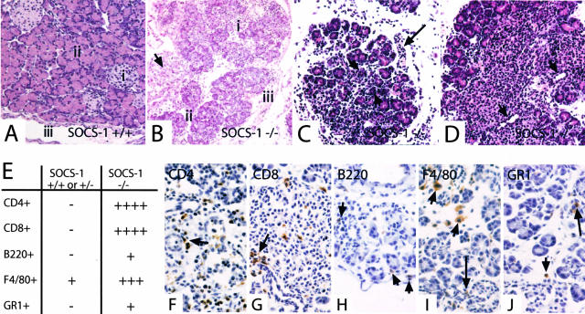
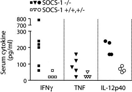
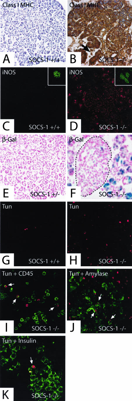
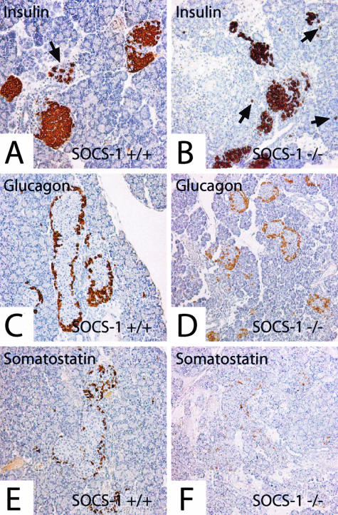
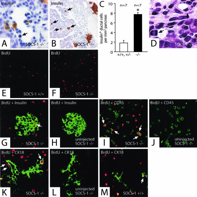
Mice with suppressor of cytokine signaling-1 (SOCS-1) deficiency die within 3 weeks of birth from a multiorgan inflammatory disease. Increased systemic levels and sensitivity of cells to the inflammatory cytokines interferon-gamma and tumor necrosis factor may contribute to the disease. Hepatitis and liver failure are thought to be the cause of the neonatal lethality in these mice. Here, we show that the pancreata of SOCS-1(-/-) mice are also severely affected by inflammation, displaying extensive edema and infiltration by T cells and macrophages. Acinar cells in particular were atrophied and reduced in their zymogen content. The expression of inflammatory markers, including class I major histocompatibility complex and inducible nitric oxide synthase, were increased in the SOCS-1(-/-) pancreas. Although there was generalized up-regulation of class I major histocompatibility complex, inducible nitric oxide synthase expression was more prominent on exocrine tissues. There appeared to be preferential damage and apoptosis of exocrine over endocrine components. Unexpectedly, increased islet neogenesis, possibly from proliferating ductal cells, was observed in the pancreas of SOCS-1(-/-) mice. This is reminiscent of the pancreatitis and islet neogenesis that occur in mice that transgenically overexpress interferon-gamma and/or tumor necrosis factor. This study suggests that in addition to liver failure, the pancreatitis may also be an important contributor to the neonatal lethality in SOCS-1(-/-) mice.
Figures
Similar articles
-
Suppressor of cytokine signaling-1 regulates the sensitivity of pancreatic beta cells to tumor necrosis factor.J Biol Chem. 2002 Aug 2;277(31):27945-52. doi: 10.1074/jbc.M110214200. Epub 2002 May 24. J Biol Chem. 2002. PMID: 12032139
-
Suppressor of cytokine signaling 3 (SOCS-3) protects beta -cells against interleukin-1beta - and interferon-gamma -mediated toxicity.Proc Natl Acad Sci U S A. 2001 Oct 9;98(21):12191-6. doi: 10.1073/pnas.211445998. Epub 2001 Oct 2. Proc Natl Acad Sci U S A. 2001. PMID: 11593036 Free PMC article.
-
Suppressor of cytokine signaling gene expression in human pancreatic islets: modulation by cytokines.Eur J Endocrinol. 2005 Mar;152(3):485-9. doi: 10.1530/eje.1.01856. Eur J Endocrinol. 2005. PMID: 15757867
-
Anti-inflammatory properties of pro-inflammatory interferon-gamma.Int Immunopharmacol. 2003 Sep;3(9):1247-55. doi: 10.1016/S1567-5769(03)00131-0. Int Immunopharmacol. 2003. PMID: 12890422 Review.
-
Acute Pancreatitis: A Multifaceted Set of Organelle and Cellular Interactions.Gastroenterology. 2019 May;156(7):1941-1950. doi: 10.1053/j.gastro.2018.11.082. Epub 2019 Jan 18. Gastroenterology. 2019. PMID: 30660726 Free PMC article. Review.
Cited by
-
MYL9 deficiency is neonatal lethal in mice due to abnormalities in the lung and the muscularis propria of the bladder and intestine.PLoS One. 2022 Jul 8;17(7):e0270820. doi: 10.1371/journal.pone.0270820. eCollection 2022. PLoS One. 2022. PMID: 35802750 Free PMC article.
-
Resolving the conundrum of islet transplantation by linking metabolic dysregulation, inflammation, and immune regulation.Endocr Rev. 2008 Aug;29(5):603-30. doi: 10.1210/er.2008-0006. Epub 2008 Jul 29. Endocr Rev. 2008. PMID: 18664617 Free PMC article. Review.
-
Carbachol protects the intestinal barrier in severe acute pancreatitis by regulating Cdc42/F-actin cytoskeleton.Exp Ther Med. 2020 Sep;20(3):2828-2837. doi: 10.3892/etm.2020.8985. Epub 2020 Jul 10. Exp Ther Med. 2020. PMID: 32765779 Free PMC article.
-
In vivo imaging of type 1 diabetes immunopathology using eye-transplanted islets in NOD mice.Diabetologia. 2019 Jul;62(7):1237-1250. doi: 10.1007/s00125-019-4879-0. Epub 2019 May 14. Diabetologia. 2019. PMID: 31087105 Free PMC article.
-
Expression of SOCS-1 in the liver tissues of chronic hepatitis B and its clinical significance.World J Gastroenterol. 2008 Jan 28;14(4):607-11. doi: 10.3748/wjg.14.607. World J Gastroenterol. 2008. PMID: 18203295 Free PMC article.
References
-
- Norman JG, Fink GW, Franz MG. Acute pancreatitis induces intrapancreatic tumor necrosis factor gene expression. Arch Surg. 1995;130:966–970. - PubMed
-
- Hunger RE, Mueller C, Z’Graggen K, Friess H, Buchler MW. Cytotoxic cells are activated in cellular infiltrates of alcoholic chronic pancreatitis. Gastroenterology. 1997;112:1656–1663. - PubMed
-
- Denham W, Yang J, Fink G, Denham D, Carter G, Ward K, Norman J. Gene targeting demonstrates additive detrimental effects of interleukin 1 and tumor necrosis factor during pancreatitis. Gastroenterology. 1997;113:1741–1746. - PubMed
-
- Grewal HP, Mohey el Din A, Gaber L, Kotb M, Gaber AO. Amelioration of the physiologic and biochemical changes of acute pancreatitis using an anti-TNF-α polyclonal antibody. Am J Surg. 1994;167:214–219. - PubMed
-
- Hughes CB, Grewal HP, Gaber LW, Kotb M, El-din AB, Mann L, Gaber AO. Anti-TNFα therapy improves survival and ameliorates the pathophysiologic sequelae in acute pancreatitis in the rat. Am J Surg. 1996;171:274–280. - PubMed
Publication types
MeSH terms
Substances
LinkOut - more resources
Full Text Sources
Medical
Molecular Biology Databases
