Comparative Study
. 2004 Sep;165(3):987-95.
doi: 10.1016/s0002-9440(10)63360-3.

Morphological characterization of Thioflavin-S-positive amyloid plaques in transgenic Alzheimer mice and effect of passive Abeta immunotherapy on their clearance

Affiliations
Comparative Study

Morphological characterization of Thioflavin-S-positive amyloid plaques in transgenic Alzheimer mice and effect of passive Abeta immunotherapy on their clearance

Thierry Bussière et al. Am J Pathol. 2004 Sep.

Abstract

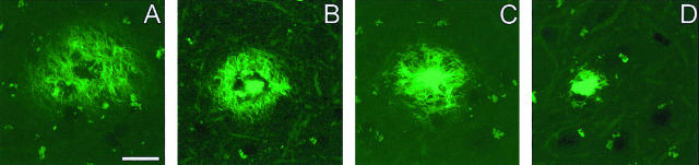
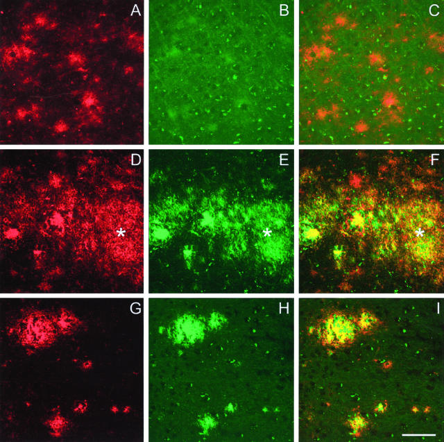
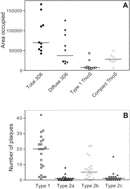
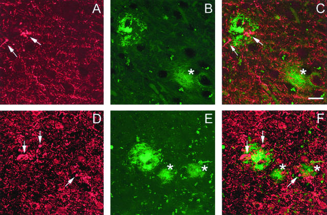
Transgenic mice mimicking certain features of Alzheimer's disease (AD)-pathology, namely amyloid plaques and neurofibrillary tangles, have been developed in an effort to better understand the mechanism leading to the formation of these characteristic cerebral lesions. More recently, these animal models have been widely used to investigate emergent therapies aimed at the reduction of the cerebral amyloid load. Several studies have shown that immunotherapy targeting the amyloid peptide (Abeta) is efficacious at clearing the amyloid plaques or preventing their formation, and at reducing the memory/behavior impairment observed in these animals. In AD, different types of plaques likely have different pathogenic significance, and further characterization of plaque pathology in the PDAPP transgenic mice would enhance the evaluation of potential therapeutics. In the present study, a morphological classification of amyloid plaques present in the brains of PDAPP mice was established by using Thioflavin-S staining. Neuritic dystrophy associated with amyloid plaques was also investigated. Finally, the efficacy of passive immunization with anti-Abeta antibodies on the clearance of Thio-S positive amyloid plaques was studied. Our results show that distinct morphological types of plaques are differentially cleared depending upon the isotype of the antibody.


Figures

Figure 1
Figure 1
Morphological classification of amyloid plaques in PDAPP mice. The classification of the amyloid plaques was established based on the morphology after Thio-S staining. A: Plaques type 1 are displayed as a mesh of stained fibrils. Compact plaques exhibit a central dense core surrounded either by a corona of fibril-like material (type 2a, B), or radiating material (type 2b, C). The most compact plaques appeared as strongly Thio-S-positive structures without any surrounding materials. They are called “burned-out” plaques, as referred to the human neuropathology classification (type 2c, D). Scale bar in (A) represents 20 μm for (A–C) and 15 μm for (D).
Figure 2
Figure 2
Double-staining 3D6/Thio-S. Top: 3D6-immunoreactive diffuse plaques (A and C) are not detected by Thio-S staining (B). Middle: Plaques with a reticular appearance that are both 3D6- and Thio-S-positive are called plaques type 1 (asterisk, D to F) to differentiate them from the diffuse plaque 3D6-immunoreactive only. Bottom: Central dense core of compact plaques type 2b are immunolabeled with 3D6 (G and I) and stained by Thio-S (H and I). Periphery of the compact plaques type 2b are 3D6-immunoreactive (G and I), but mainly Thio-S-negative (I). Scale bar, 20 μm.
Figure 3
Figure 3
Different types of plaques in PDAPP mice. A: Thio-S-positive plaques represent a subset of the 3D6-immunoreactive plaques. Sections from 10 control animals were immunostained with 3D6 before being stained with Thio-S, and three fields per section were imaged by confocal microscopy. Each dot represents the median value of the area occupied for one animal. For each animal, total area occupied by all plaques 3D6-immunoreactive, area occupied only by diffuse plaques 3D6-immunoreactive, area occupied by type 1 plaque Thio-S-positive, and area occupied by compact plaque Thio-S-positive were determined with NIH Image 1.63. B: Relative proportion of the different types of plaques in control PDAPP mice. The number of plaques in control groups from two immunization studies were pooled together. Each dot represents the number of plaques for a given animal.
Figure 4
Figure 4
Neuritic lesions associated with plaques in PDAPP mice. Top: Swollen or distorted neurites immunoreactive with SMI 312 against phosphorylated neurofilaments (arrows in A and C) are associated with a compact plaque type 2a (B and C). A Thio-S-positive plaque type 1 in the same section (asterisk in B and C) was devoid of such associated lesion. Bottom: Clusters of dystrophic neurites (arrows in D and F) are localized in the immediate vicinity of a compact plaque type 2b (E and F). Very few neuritic lesions were associated with plaques type 1 (asterisks in E and F). The weaker SMI312 immunostaining observed in the top panel was because of the localization of the microscopic field, where a lower density of processes was observed. Scale bar in (C) represents 20 μm for (A–C) and 30 μm for (D–F).
Figure 5
Figure 5
Effect of passive immunization on removal of different types of plaques. Anti Aβ-antibodies of different isotypes have differential efficacy on the clearance of amyloid plaques. IgG2a antibody (12B4) was efficacious in significantly reducing both type 1 and compact plaques type 2b (P = 0.001 and 0.007, respectively). IgG1 (10D5) and IgG2b (12A11) antibodies had no significant effect on clearance of Thio-S-positive plaques, and were not significantly different from PBS-treated age-matched PDAPP control group.

References

    1. Mirra SS, Gearing M, Nash F. Neuropathologic assessment of Alzheimer’s disease. Neurology. 1997;49:S14–S16. - PubMed
    1. Delaère P, Duyckaerts C, He Y, Piette F, Hauw JJ. Subtypes and differential laminar distributions of beta A4 deposits in Alzheimer’s disease: relationship with the intellectual status of 26 cases. Acta Neuropathol. 1991;81:328–335. - PubMed
    1. Dickson DW. The pathogenesis of senile plaques. J Neuropathol Exp Neurol. 1997;56:321–339. - PubMed
    1. Knowles RB, Gomez-Isla T, Hyman BT. Abeta associated neuropil changes: correlation with neuronal loss and dementia. J Neuropathol Exp Neurol. 1998;57:1122–1130. - PubMed
    1. Hsiao K, Chapman P, Nilsen S, Eckman C, Harigaya Y, Younkin S, Yang F, Cole G. Correlative memory deficits, Abeta elevation, and amyloid plaques in transgenic mice. Science. 1996;274:99–102. - PubMed

Publication types

MeSH terms