Comparative Study
. 2004 Sep;165(3):1033-44.
doi: 10.1016/S0002-9440(10)63364-0.

Heterozygous inactivation of the vinculin gene predisposes to stress-induced cardiomyopathy

Affiliations
Comparative Study

Heterozygous inactivation of the vinculin gene predisposes to stress-induced cardiomyopathy

Alice E Zemljic-Harpf et al. Am J Pathol. 2004 Sep.

Abstract

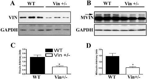
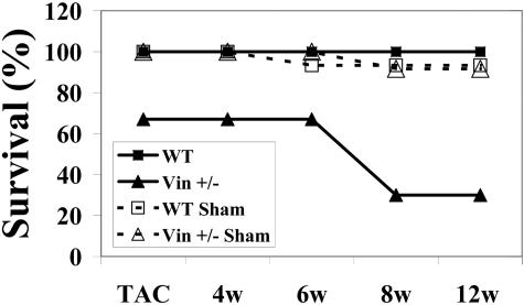
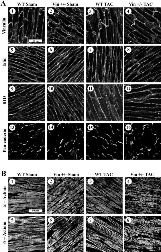
Vinculin and its muscle splice variant metavinculin link focal adhesions and cell-to-cell contact sites to the actin cytoskeleton. We hypothesized that normal expression of vinculin isoforms would be essential for integrity of cardiomyocytes and preservation of normal cardiac function. We studied heterozygous vinculin knockout mice (Vin+/-) that develop and breed normally. The Vin+/- mice displayed: 1) a 58% reduction of vinculin and a 63% reduction of metavinculin protein levels versus wild-type littermates; 2) normal basal cardiac function and histology but abnormal electrocardiograms, intercalated disks, and ICD-related protein distribution; 3) increased mortality following acute hemodynamic stress imposed by transverse aortic constriction (TAC); 4) cardiac dysfunction by 6 weeks post-TAC; and 5) misalignment of alpha-actinin containing Z-lines and abnormal myocardial ultrastructure despite preserved cardiac function. Decreased expression of vinculin/metavinculin leads to abnormal myocyte structure without baseline physiological evidence of cardiac dysfunction. These structural changes predispose to stress-induced cardiomyopathy.


Figures

Figure 1
Figure 1
Vinculin and metavinculin protein levels are reduced in Vin+/− hearts. Whole heart lysates were analyzed by Western blotting simultaneously with antibodies to vinculin/metavinculin and GAPDH. Representative experiments are shown. A: Vin/GAPDH Western blot. B: Mvin/GAPDH Western blot. C and D: Densitometric analysis performed by normalizing vinculin or metavinculin expression to simultaneously measured GAPDH. The mean intensity per group is shown in arbitrary units. (n = 6, each group, *P < 0.003 vs. WT).
Figure 2
Figure 2
Basal cardiac function and morphology is normal in Vin+/− mice. No significant differences were detected in Vin+/− vs. WT mice when evaluated by histological analysis or closed-chest cardiac catheterization at baseline and during dobutamine infusion. (Cardiac catheterization: WT n = 4 and Vin+/− n = 5). A and B: Myocardial sections stained with hematoxylin and eosin from 3-month-old wild-type (A) and Vin+/− (B) mice revealed normal myocyte architecture and lack of fibrosis in both groups. (Magnification, ×400). C: Heart rate. D: LV systolic pressure. E: LV dP/dtmax. F: LV dP/dt min.
Figure 3
Figure 3
Electrocardiograms display widened QRS complexes in Vin+/− mice. No significant differences in heart rate or PR interval duration were seen between WT and Vin+/− mice, whereas QRS duration (measured as indicated by the arrows) was significantly prolonged in Vin+/− animals (n = 12 per group, *P < 0.007). A: Heart rate in WT and Vin+/− mice. B: PR duration (ms) in WT and Vin+/− mice. C: QRS duration (ms) in WT and Vin+/− mice. D and E: Representative ECG tracings in WT and Vin+/− mice, respectively.
Figure 4
Figure 4
Altered intercalated disks are detected in Vin+/− hearts as visualized by immunomicroscopy of vinculin, connexin 43, and cadherin. Immunofluorescent staining with an anti-vinculin antibody in hearts of 4-month-old WT (A) and Vin+/− (B) mice. Vin+/− hearts showed normal costamere distribution but unorganized intercalated disk appearance via vinculin staining. Identical findings were detected with anti-Cx43 and anti-cadherin antibodies. Scoring for ICD disorganization was performed by two observers in a genotype-blinded fashion. TRITC-labeled phalloidin was used to visualize F-actin and DAPI for nuclei. A and B: Vinculin staining in WT (A) and Vin+/− (B). C and D: Cx43 (green), F-actin (red), and DAPI (blue) staining in WT (C) and Vin+/− (D). E and F: Cadherin (green) and F-actin (red) staining in WT (E) and Vin+/− (F). G: Graphical representation of ICD scoring in WT and Vin+/− hearts. ICD structure was judged by their Cx43 and cadherin staining pattern as either normal or wide. Percentage of wide ICDs is shown for each genotype (*P < 0.001 Vin+/− vs. WT, #P < 0.002 Vin+/− vs. WT).
Figure 5
Figure 5
Vin+/− mice show increased mortality following pressure overload. Transverse aortic constriction was performed in nine Vin+/− mice and 33% (n = 3) died within 6 hours, following initial postoperative recovery. The remaining six Vin+/− mice and all other TAC and sham animals from both genotypes were then followed for up to 12 weeks after surgery. Surviving animals at the following specified time points: TAC (0w): WT Sham, (n = 7), Vin+/− Sham (n = 6), WT TAC (n = 9), Vin+/− TAC (n = 6); 4 weeks post-TAC (4w): WT Sham, (n = 7), Vin+/− Sham (n = 6), WT TAC (n = 9), Vin+/− TAC (n = 6); 6 weeks post-TAC (6w): WT Sham, (n = 6), Vin+/− Sham (n = 6), WT TAC (n = 9), Vin+/− TAC (n = 6); 8 weeks post-TAC (8w): WT Sham, (n = 6), Vin+/− Sham (n = 5), WT TAC (n = 9), Vin+/− TAC (n = 3); 12 weeks post-TAC (12w): WT Sham, (n = 6), Vin+/− Sham (n = 5), WT TAC (n = 9), Vin+/− TAC (n = 3)
Figure 6
Figure 6
Echocardiographic analysis following chronic pressure overload showed that LV function became compromised beginning at 6 weeks post-TAC only in Vin+/− mice. Animals were evaluated by echocardiography at baseline (BL) and subsequently at 4, 6, 8, and 12 weeks post-TAC. A: Left ventricular fractional shortening (LV %FS). B: Circumferential fiber shortening (Vcf). C: Left ventricular end-diastolic posterior wall thickness (LVPWTd). D: Left ventricular end-diastolic interventricular septum thickness (IVSTd). E: Left ventricular end-diastolic diameter (LVEDD). F: Left ventricular end systolic diameter (LVESD). Baseline: WT (n = 11), Vin+/− (n = 12). 4 weeks (4w): WT-Sham (n = 5), WT-TAC (n = 6), Vin+/− Sham (n = 6), Vin+/− TAC (n = 6). 6 weeks (6w): WT-Sham (n = 5), WT-TAC (n = 6), Vin+/− Sham (n = 4), Vin+/− TAC (n = 6). 8 weeks (8w): WT-Sham (n = 5), WT-TAC (n = 7), Vin+/− Sham (n = 5), Vin+/− TAC (n = 6). 12 weeks (12w): WT-Sham (n = 4), WT-TAC (n = 7), Vin+/− Sham (n = 5), Vin+/− TAC (n = 3). P values: LV %FS: *WT TAC vs. Vin+/− TAC, P < 0.004. §Vin+/− TAC vs. Vin+/− Sham, P < 0.02. Vcf: *WT TAC vs. Vin+/− TAC, P < 0.01. §Vin+/− TAC vs. Vin+/− Sham, P < 0.03. LVPWTd: §Vin+/− TAC vs. Vin+/− Sham, P < 0.005. # WT TAC vs. WT Sham, P < 0.02. IVSd: §Vin+/− Sham vs. Vin+/− TAC, P < 0.001. #WT Sham vs. WT TAC, P < 0.003. LVESD *WT TAC vs. Vin+/− TAC, P < 0.002. §Vin+/− TAC vs. Vin+/− Sham P < 0.03. #WT TAC vs. WT Sham, P < 0.02.
Figure 7
Figure 7
Z-lines are disorganized in myocardial tissue from Vin+/− mice. Confocal microscopic analysis of vinculin and associated proteins was performed at 4 weeks after surgery in all groups (n = 3 per group), a time point when no physiological differences were detected in Vin+/− vs. WT animals. A: Immunostaining for vinculin (1–4) and the associated proteins, talin, (5-8), β1D-integrin (9–12), and pan-cadherin (13–16). Abnormal intercalated disk structure is evident in Vin+/− animals not WT controls as indicated by vinculin and cadherin distribution. Localization of talin and B1D were similar in all groups. B: Immunostaining for the vinculin-binding protein α-actinin (1–8). WT sham hearts (1 and 5) displayed normal Z-line structure as compared to Vin+/− sham hearts (2 and 6) which showed mild irregularities in Z-lines. Normal parallel Z-line staining continued to be seen in WT-TAC hearts (3 and 7) while the Vin+/− TAC Z-lines (4 and 8) became even more irregularly distributed. Magnified micrographs from 1 to 4 are shown below each in panels 5–8.
Figure 8
Figure 8
Transmission electron microscopy of left ventricular mouse myocardium. Cardiac muscle from wild-type mice (A) shows well aligned arrays and insertions of myofibrils at Z-lines and intercalated disks. Mitochondria are visible in packed strands and are of normal size. Myofibrils of Vin+/− myocardial tissue (B) appear separated from the ICDs (arrows) as well as from Z-lines (arrowheads). Myofibrils in Vin+/− appear less densely packed than in WT muscle and were interrupted occasionally by swollen and disorganized mitochondria. C and D: Higher magnification views of A and B. Panels are representative of micrographs obtained from three animals of each genotype.

References

    1. Rudiger M, Korneeva N, Schwienbacher C, Weiss EE, Jockusch BM. Differential actin organization by vinculin isoforms: implications for cell type-specific microfilament anchorage. FEBS Lett. 1998;431:49–54. - PubMed
    1. Schlaepfer DD, Hunter T. Signal transduction from the extracellular matrix—a role for the focal adhesion protein-tyrosine kinase FAK. Cell Struct Funct. 1996;21:445–450. - PubMed
    1. Hildebrand JD, Schaller MD, Parsons JT. Identification of sequences required for the efficient localization of the focal adhesion kinase, pp125FAK, to cellular focal adhesions. J Cell Biol. 1993;123:993–1005. - PMC - PubMed
    1. Lu MH, DiLullo C, Schultheiss T, Holtzer S, Murray JM, Choi J, Fischman DA, Holtzer H. The vinculin/sarcomeric-α-actinin/α-actin nexus in cultured cardiac myocytes. J Cell Biol. 1992;117:1007–1022. - PMC - PubMed
    1. Jockusch BM, Isenberg G. Vinculin and α-actinin: interaction with actin and effect on microfilament network formation. Cold Spring Harbor Symp Quant Biol. 1982;46 Pt 2:613–623. - PubMed

Publication types