. 2004 Nov 15;561(Pt 1):65-90.
doi: 10.1113/jphysiol.2004.073353. Epub 2004 Aug 26.

Anatomical, physiological and molecular properties of Martinotti cells in the somatosensory cortex of the juvenile rat

Affiliations

Anatomical, physiological and molecular properties of Martinotti cells in the somatosensory cortex of the juvenile rat

Yun Wang et al. J Physiol. .

Abstract

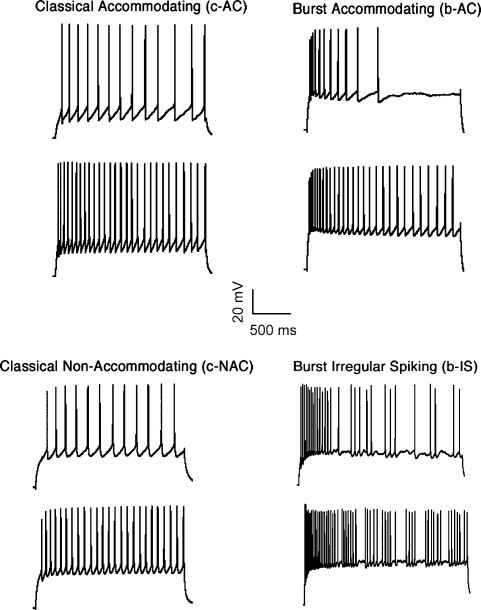
Whole-cell patch-clamp recordings followed by histochemical staining and single-cell RT-PCR were obtained from 180 Martinotti interneurones located in layers II to VI of the somatosensory cortex of Wistar rats (P13-P16) in order to examine their anatomical, electrophysiological and molecular properties. Martinotti cells (MCs) mostly displayed ovoid-shaped somata, bitufted dendritic morphologies, and axons with characteristic spiny boutons projecting to layer I and spreading horizontally across neighbouring columns more than 1 mm. Electron microscopic examination of MC boutons revealed that all synapses were symmetrical and most synapses (71%) were formed onto dendritic shafts. MCs were found to contact tuft, apical and basal dendrites in multiple neocortical layers: layer II/III MCs targeted mostly layer I and to a lesser degree layer II/III; layer IV MCs targeted mostly layer IV and to a lesser degree layer I; layer V and VI MCs targeted mostly layer IV and layer I and to a lesser degree the layer in which their somata was located. MCs typically displayed spike train accommodation (90%; n = 127) in response to depolarizing somatic current injections, but some displayed non-accommodating (8%) and a few displayed irregular spiking responses (2%). Some accommodating and irregular spiking MCs also responded initially with bursts (17%). Accommodating responses were found in all layers, non-accommodating mostly in upper layers and bursting mostly in layer V. Single-cell multiplex RT-PCR performed on 63 MCs located throughout layers II-VI, revealed that all MCs were somatostatin (SOM) positive, and negative for parvalbumin (PV) as well as vasoactive intestinal peptide (VIP). Calbindin (CB), calretinin (CR), neuropeptide Y (NPY) and cholecystokinin (CCK) were co- expressed with SOM in some MCs. Some layer-specific trends seem to exist. Finally, 24 accommodating MCs were examined for the expression of 26 ion channel genes. The ion channels with the highest expression in these MCs were (from highest to lowest); Cabeta1, Kv3.3, HCN4, Cabeta4, Kv3.2, Kv3.1, Kv2.1, HCN3, Caalpha1G, Kv3.4, Kv4.2, Kv1.1 and HCN2. In summary, this study provides the first detailed analysis of the anatomical, electrophysiological and molecular properties of Martinotti cells located in different neocortical layers. It is proposed that MCs are crucial interneurones for feedback inhibition in and between neocortical layers and columns.


Figures

Figure 1
Figure 1. Features of a layer V MC
A, 3-D computer reconstruction (soma and dendrites are in red, axon in blue, boutons marked with dots). Note bitufted morphology, ovoid somata, axon targeting layers I, IV and V and downward dendritic projections. B, photo of spiny boutons on MC axons (arrows). C, EM image of MC boutons in layer I. D, classical accommodating discharge to sustained somatic current injections. E, single-cell RT-PCR results: agarose gel showing the expression of mRNAs encoding for the CaBPs (calbindin, CB; parvalbumin, PV; and calretinin, CR); the neuropeptides (neuropeptide Y, NPY; vasoactive intestinal peptide, VIP; somatostatin, SOM; cholecystokinin, CCK); the voltage-activated K+ channels (Kv1.1, Kv1.2, Kv1.4, Kv1.6, Kv2.1, Kv2.2, Kv3.1, Kv3.2, Kv3.3, Kv3.4, Kv4.2 and Kv4.3) and their auxiliary subunits (Kvβ1 and Kvβ2); the K+/Na+ permeable hyperpolarization-activated ion channels (HCN1, HCN2, HCN3 and HCN4); the Ca2+-activated K+ channel (SK2); the voltage-activated calcium channels (Caα1A, Caα1B, Caα1G and Caα1I) and their auxiliary subunits (Caβ1, Caβ3 and Caβ4) and the ubiquitous protein GAPDH. See Table 1 for the lists of the primer pairs included into the different multiplexes, the name and accession number of the genes amplified and the length of the PCR product.
Figure 6
Figure 6. Major electrophysiological parameters for active and passive properties of MCs
Example responses to different stimulation protocols used for electrophysiological characterization. AP Waveform, APs evoked by brief step current injections. AP Threshold, the threshold for AP discharge was measured during a ramp current injection. AP Drop, changes in AP amplitude during a discharge train evoked by a step current injection. IV, the current–voltage relationship was extracted by a series of subthreshold current injections. Delta, the response to a brief (1 ms) hyperpolarizing current was used to measure the membrane time constant. Sag, the response to a hyperpolarizing current step was analysed to reveal the existence of hyperpolarization-dependent inward currents. s-AHP, the existence and degree of slow membrane hyperpolarization following rapid discharge. ID Rest, discharge response to increasing current injections was used for assessing the current-discharge properties of MCs. ID Threshold, discharge response to near-threshold current injections, used for neuronal classification by revealing the existence of onset delays or bursts.
Figure 2
Figure 2. Morphometric analysis of a layer V MC
A, diagram of a reconstructed neurone and demonstration of its morphometric parameters (soma and dendrites in red, axon in blue). ASD, axon Sholl distance. Serial Sholl circles with 20 μm-stepped radii were centred in the soma. Numbers of intersections within each Sholl circle were counted and graphed as a function of distance to the centre. ASL, axonal segment length; defined as the length of axonal segment between two neighbouring branch points or between a branch point and an end point. ABO, axonal branch order; represents the frequency of axonal branching, its increases after each branch point from the initial axon segment. MABA or MDBA, maximum axonal or dendritic branch angle; the maximum angle formed between the extending distal line of a parent axonal segment and the daughter axonal segments (the lower bigger angle marked with a dash arc). PABA or PDBA, planar axonal or dendritic branching angle; the maximum angle formed between the extending distal line of the parent axonal segment and a daughter axonal segment (two planar angles were formed after branching). B, histograms of five major axonal parameters (for the layer V MC in A). The ASD histogram shows a first peak due to the axonal cluster formed in layer IV, and a second peak about 600–700 μm away due to the dense axonal cluster in layer I. The ASL histogram presents the different lengths of axonal segments. The ABO histogram presents distributions of axonal segment distribution in terms of branch orders. Most segments are 6–19th branch order. The MABA histogram shows the axonal segment distribution of this MC. The MABAs vary from less than 10 deg to nearly 180 deg, but most MABAs are arranged between 40 and 100 deg. The PABA histogram at the bottom shows the axonal segment distribution plotted according to planar axonal branch angles. C, histograms of five major dendritic parameters (for the layer V MC in A) as before for axons.
Figure 3
Figure 3. 3-D computer reconstructions of MCs in different layers
A1, layer II/III MC with a prominent axonal cluster in layer I and only a few collaterals ramified around its soma, and dendrites that extend vertically. A2, ASD distribution of layer II/III MCs displays two peaks. The first peak (around 100 μm) reflects the axonal cluster around soma. B1, layer IV MC with a prominent axonal cluster around the soma and sparse cluster in layer I, and localized dendrites. B2, ASD distribution of layer IV MCs. Only one peak was formed close to the soma. C1, layer V MC with a prominent axonal cluster in layers IV and I, a sparse cluster around the soma, and vertically extending dendrites. D, layer VI MC. This MC has a dendritic tree localized in the infragranular layers and an axonal tree forming clusters in layer I, IV and VI. C2, ASD distribution of layer V MCs also display two peaks.
Figure 4
Figure 4. Random EM examination of MC boutons
A, MC boutons targeting a dendritic shaft of a putative PC. The synapse is pointed out with an unfilled wide arrow. Note the transmitter vesicles in the filled bouton and the postsynaptic shaft with light cytoplasm on the right. B, MC bouton targeting a dendritic shaft of a putative interneurone. Note the postsynaptic shaft with more cell organelles and darker cytoplasm. C, axo-spinous synapse. Note rigid and parallel pre-postsynaptic membrane appositions. D, axo-somatic synapse. Note synaptic vesicles along the presynaptic membrane and two active zones (rigid and parallel pre-postsynaptic membrane segments) indicated by two arrows. Nuc, nucleus; b, bouton; sh, dendritic shaft; sp, spine; arrow, synaptic junction; all scale bars, 0.5 μm.
Figure 5
Figure 5. Four different discharge responses found in MCs
The discharge responses were induced by sustained somatic current injections. Top, accommodating (AC) responses show gradual increase in interspike intervals, including classical (c-AC, regular firing onset; top left) and bursting (b-AC, burst firing onset; top right). Bottom left, classical non-accommodating (c-NAC) cells display virtually no change in inter-spike intervals with regular firing onset. Bottom right, burst irregular spiking (b-IS) cells display abrupt changes in inter-spike intervals during steady state and bursting onset.
Figure 7
Figure 7. Representative samples of electrophysiological parameters with the highest and lowest coefficient of variation
A, AP waveform traces. Some of the AP parameters showed the lowest coefficient of variation. B, AP drop traces. Some of the AP drop parameters showed the highest coefficient of variation. C, graph describing the absolute CVs for different electrophysiological parameters sorted from the less to the most variable. See Table 5 for the identity of the electrophysiological parameters.
Figure 8
Figure 8. Gene expression in MCs
A, 3-D computer reconstruction. B, classical accommodation responses in a layer V MC. C, chart showing the percentage of MCs expressing different mRNAs encoding for the CaBPs (calbindin, CB; parvalbumin, PV; and calretinin, CR), neuropeptides (neuropeptide Y, NPY; vasoactive intestinal peptide, VIP; somatostatin, SOM; cholecystokinin, CCK) sorted according to layer. D, chart showing percentages of c-AC MCs expressing different mRNAs encoding for the voltage-activated K+ channels (Kv1.1, Kv1.2, Kv1.4, Kv1.6, Kv2.1, Kv2.2, Kv3.1, Kv3.2, Kv3.3, Kv3.4, Kv4.2 and Kv4.3) and their auxiliary subunits (Kvβ1 and Kvβ2); the K+/Na+ permeable hyperpolarization-activated ion channels (HCN1, HCN2, HCN3 and HCN4) the Ca2+-activated K+ channel SK2; and the voltage-activated calcium channels (Caα1A, Caα1B, Caα1G and Caα1I) and their auxiliary subunits (Caβ1, Caβ3 and Caβ4).

References

    1. Armstrong DM, LeRoy S, Shields D, Terry RD. Somatostatin-like immunoreactivity within neuritic plaques. Brain Res. 1985;338:71–79. 10.1016/0006-8993(85)90249-5. - DOI - PubMed
    1. Beal MF, Clevens RA, Mazurek MF. Somatostatin and neuropeptide Y immunoreactivity in Parkinson's disease dementia with Alzheimer's changes. Synapse. 1988;2:463–467. - PubMed
    1. Bernander O, Douglas RJ, Martin KA, Koch C. Synaptic background activity influences spatiotemporal integration in single pyramidal cells. Proc Natl Acad Sci U S A. 1991;88:11569–11573. - PMC - PubMed
    1. Bernander O, Koch C, Douglas RJ. Amplification and linearization of distal synaptic input to cortical pyramidal cells. J Neurophysiol. 1994;72:2743–2753. - PubMed
    1. Cauli B, Audinat E, Lambolez B, Angulo MC, Ropert N, Tsuzuki K, Hestrin S, Rossier J. Molecular and physiological diversity of cortical non pyramidal cells. J Neurosci. 1997;17:3894–3906. - PMC - PubMed