. 2004 Sep;78(18):9633-44.
doi: 10.1128/JVI.78.18.9633-9644.2004.

Autographa californica multiple nucleopolyhedrovirus exon0 (orf141), which encodes a RING finger protein, is required for efficient production of budded virus

Affiliations

Autographa californica multiple nucleopolyhedrovirus exon0 (orf141), which encodes a RING finger protein, is required for efficient production of budded virus

Xiaojiang Dai et al. J Virol. 2004 Sep.

Abstract

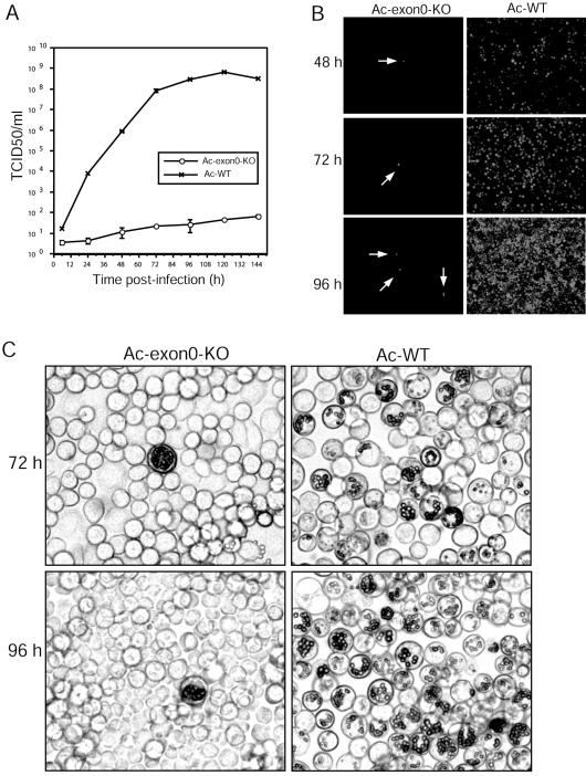
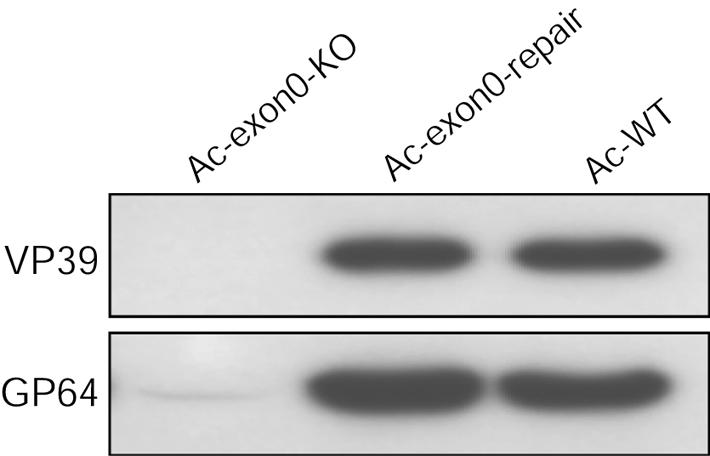
exon0 (orf141) of Autographa californica multiple nucleopolyhedrovirus (AcMNPV) is a highly conserved baculovirus gene that codes for a predicted 261-amino-acid protein. Located in the C-terminal region of EXON0 are a predicted leucine-rich coiled-coil domain and a RING finger motif. The 5' 114 nucleotides of exon0 form part of ie0, which is a spliced gene expressed at very early times postinfection, but transcriptional analysis revealed that exon0 is transcribed as a late gene. To determine the role of exon0 in the baculovirus life cycle, we used AcMNPV bacmids and generated exon0 knockout viruses (Ac-exon0-KO) by recombination in Escherichia coli. Ac-exon0-KO progressed through the very late phases in Sf9 cells, as evidenced by the development of occlusion bodies in the nuclei of the transfected or infected cells. However, production of budded virus (BV) in Ac-exon0-KO-infected cells was reduced at least 3 orders of magnitude in comparison to that in wild-type virus infection. Microscopy revealed that Ac-exon0-KO was restricted primarily to the cells initially infected, exhibiting a single-cell infection phenotype. Slot blot assays and Western blot analysis indicated that exon0 deletion did not affect the onset or levels of viral DNA replication or the expression of IE1, IE0, and GP64 prior to BV release. These results demonstrate that exon0 is required for efficient production of BV in the AcMNPV life cycle but does not affect late occlusion-derived virus.


Figures

FIG. 1.
FIG. 1.
(A) Schematic map of the exon0-ie0-ie1 gene region showing the transcription and splicing pattern of ie1 and ie0 and the relative positions and orientations of the exon0 (orf141), ie1, ie0, me53, orf142, orf143, orf144, orf145, orf146, and odv-e56 ORFs. The ie0 intron and transcript are indicated as a dotted line and solid line respectively, above the ORFs. The resulting spliced ORF of IE0 is shown indicating the regions that originate from exon0 (black), ie1 UTR (white), and ie1 (grey). On the basis of the locations of polyadenylation signals, potential exon0 transcripts are indicated below the ORFs. Solid arrows agree with the observed sizes on Northern blots. (B) Northern blot analysis of exon0 transcription in AcMNPV-infected Sf9 cells from 3 to 72 h p.i. The location of the strand-specific probe is shown above the exon0 ORF in panel A (black line). Numbers above each lane indicate the time (hours) p.i. when RNA was isolated. The sizes of exon0 RNAs are indicated on the right. (C) 5′ RACE analysis of the exon0 transcriptional start site. Alignment of the ie0-exon0 promoter with the sequence obtained with exon0-specific primers (GSP-2A, GSP-2B). The TATA box (TATAAA), early transcriptional start site (CAGT), and late promoter (ATAAG) are shown in bold, and the translation start codon is underlined. The arrowhead shows the location of the transcription initiation site of exon0.
FIG. 2.
FIG. 2.
Alignment of EXON0 homologues from 13 baculovirus NPVs. The alignment was performed with the ClustalW algorithm (53). Identical and conserved amino acids are boxed, and regions with greater than 70% identity are shaded. Conserved cysteine and tyrosine residues in the predicted RING finger motif at the C terminus are indicated by arrowheads. The consensus RING finger motif is shown below the alignment (10). The predicted leucine-rich coiled coil and the amino acids spliced to IE1 to form IE0 are indicated by black lines above the alignment.
FIG. 3.
FIG. 3.
Construction of exon0 knockout, repair, and WT AcMNPV bacmids. (A) Schematic diagram of the transfer vector, Ac-exon0-KO, used to generate the exon0 knockout bacmid by recombination in E. coli. pAc-exon0-KO contains the zeocin resistance gene under the control of the EM7 promoter, 1,547 bp of the upstream exon0 flanking region, and 543 bp of the downstream exon0 flanking region. This leaves the ie0 splice site and the orf142 promoter region intact. (B) Schematic diagram of three viruses, Ac-exon0-KO, Ac-exon0-repair, and Ac-WT, showing the genes inserted into the polyhedrin locus by Tn7-mediated transposition. polh, polyhedrin; gfp, green fluorescent protein. (C) Positions of primer pairs used in the analysis of the WT locus and exon0 knockout locus to confirm the deletion of the exon0 ORF and correct insertion of the zeocin resistance gene cassette. Primers are indicated by arrows designated A through D. (D) Confirmation by PCR analysis of the presence or absence of sequence modifications in Ac-exon0-KO, Ac-exon0-repair, and Ac-WT. The virus analyzed is shown above each lane, and the primer pairs used are shown below.
FIG. 4.
FIG. 4.
Analysis of viral replication by the exon0 knockout virus in bacmid DNA-transfected Sf9 cells. (A) Virus growth curves of Ac-exon0-KO, Ac-exon0-repair, and Ac-BAC-WT in Sf9 cells. Cells (2.0 × 10⁶) were transfected with 2.0 μg of DNA of each virus. Cells were harvested at the indicated time points p.i., and the cell culture supernatants were harvested and assayed for the production of infectious virus by TCID50 assay. Each datum point represents the average titer derived from two independent TCID50 assays. Error bars represent standard errors. (B) Fluorescence microscopy of Ac-exon0-KO-, Ac-exon0-repair-, and Ac-WT-transfected Sf9 cells at 18 and 48 h p.t. (C) Light microscopy of Ac-exon0-KO-, Ac-exon0-repair-, and Ac-WT-transfected cells at 48, 72, and 96 h p.t.
FIG. 5.
FIG. 5.
Analysis of viral replication by the exon0 knockout virus in BV-infected Sf9 cells. (A) Virus growth curves of Ac-exon0-KO and Ac-WT in Sf9 cells. 2.0 × 10⁶ cells were infected at an MOI of 0.0002 from each virus, and supernatants were harvested at the indicated time points p.i. and assayed for the production of infectious virus by TCID50 assay. Each datum point represents the average titer derived from two independent TCID50 assays. Error bars represent standard errors. (B) Fluorescence microscopy of Ac-exon0-KO- and Ac-WT-infected Sf9 cells at 48, 72, and 96 h p.t. The single or few cells infected by Ac-exon0-KO are indicated by the arrows. (C) Light microscopy of Ac-exon0-KO- and Ac-WT-infected cells at 72 and 96 h p.t.
FIG. 6.
FIG. 6.
Plaque assay analysis of Ac-exon0-KO and Ac-WT in infected Sf9 cells. (A) Fluorescence microscopic analysis of plaques formed by Ac-exon0-KO- and Ac-WT-infected Sf9 cells at 5 days p.i. demonstrating the inability of Ac-exon0-KO to spread the infection via BV. The single or few cells infected by Ac-exon0-KO are indicated by the arrows. (B) Fluorescence microscopy of the types of infected-cell foci that contain single or few cells that are observed in Ac-exon0-KO-infected cells.
FIG. 7.
FIG. 7.
Western blot analysis of purified BV particles. Viral particles were purified from supernatants of Sf9 cells that had been transfected with Ac-exon0-KO, Ac-exon0-repair, or Ac-WT. Samples were analyzed with two monoclonal antibodies specific for nucleocapsid protein VP39 and BV-specific protein GP64.
FIG. 8.
FIG. 8.
Slot blot analysis of viral DNA replication. (A) Sf9 cells (2.0 × 10⁶) were transfected with 2.0 μg of Ac-exon0-KO, Ac-exon0-repair, or Ac-WT DNA. At the designated times p.t., cells were harvested and cell lysates were prepared for slot blot analysis. The EcoRI-T fragment of AcMNPV was labeled with [32P]dCTP and used as a hybridization probe. On the left are the times p.t. (B) Quantitative analysis of viral DNA replication by slot blot analysis. DNA was quantified by PhosphorImager analysis, and each datum point represents the average from two independent transfections. Error bars represent the standard errors.
FIG. 9.
FIG. 9.
Western blot time course analysis of IE1, IE0, and GP64 synthesis in Sf9 cells that have been transfected with Ac-exon0-KO, Ac-exon0-repair, or Ac-WT. IE1 and IE0 (A) and GP64 (B) were analyzed with monoclonal antibodies specific to each protein. Sf9 cells (2.0 × 10⁶) were transfected with 2.0 μg of DNA of each virus. At the designated times p.t., cells were harvested and cell lysates were prepared for Western blot analysis. The locations of the IE0- and IE1-specific bands are indicated on the right.

Similar articles

Cited by

References

    1. Albert, T. K., H. Hanzawa, Y. I. Legtenberg, M. J. de Ruwe, F. A. van den Heuvel, M. A. Collart, R. Boelens, and H. T. Timmers. 2002. Identification of a ubiquitin-protein ligase subunit within the CCR4-NOT transcription repressor complex. EMBO J. 21:355-364. - PMC - PubMed
    1. Albert, T. K., M. Lemaire, N. L. van Berkum, R. Gentz, M. A. Collart, and H. T. Timmers. 2000. Isolation and characterization of human orthologs of yeast CCR4-NOT complex subunits. Nucleic Acids Res. 28:809-817. - PMC - PubMed
    1. Ayres, M. D., S. C. Howard, J. Kuzio, M. Lopez-Ferber, and R. D. Possee. 1994. The complete DNA sequence of Autographa californica nuclear polyhedrosis virus. Virology 202:586-605. - PubMed
    1. Blissard, G. W. 1996. Baculovirus-insect cell interactions. Cytotechnology 20:73-93. - PubMed
    1. Blissard, G. W., and G. F. Rohrmann. 1989. Location, sequence, transcriptional mapping, and temporal expression of the gp64 envelope glycoprotein gene of the Orgyia pseudotsugata multicapsid nuclear polyhedrosis virus. Virology 170:537-555. - PubMed

Publication types

MeSH terms