. 2004 Sep;78(18):9750-62.
doi: 10.1128/JVI.78.18.9750-9762.2004.

Human cytomegalovirus UL76 encodes a novel virion-associated protein that is able to inhibit viral replication

Affiliations

Human cytomegalovirus UL76 encodes a novel virion-associated protein that is able to inhibit viral replication

Shang-Kwei Wang et al. J Virol. 2004 Sep.

Abstract

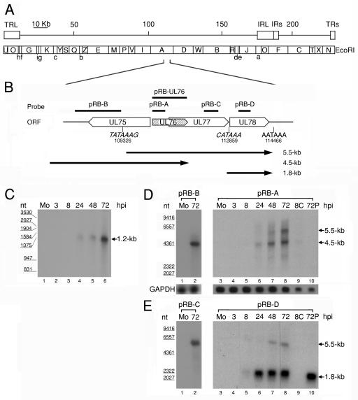
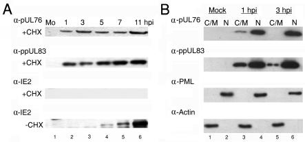
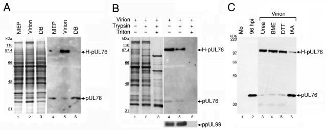
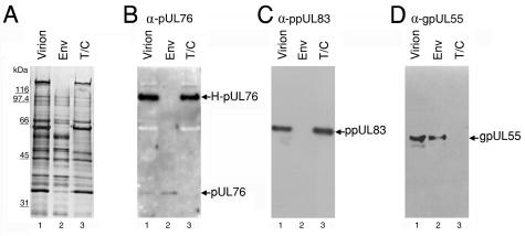
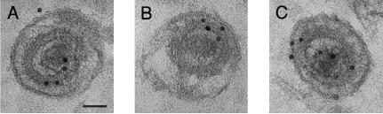
The human cytomegalovirus (HCMV) UL76 gene encodes a highly conserved herpesvirus protein, pUL76, which is able to modulate gene expression in either activation or repression. In this study, two specific transcripts were found to contain the reading frame of UL76, one a 4.5-kb and the other a 5.5-kb tricistronic mRNA encoding the UL76, UL77, and UL78 open reading frames. Both transcripts were expressed with true late kinetics, as revealed by data showing inhibition of production in the presence of phosphonoformic acid. Immediately after viral infection, pUL76 was found in the nuclear fraction and was detected in cells in the presence of the protein synthesis inhibitor cycloheximide. Subsequent virus particle purification and Western blot analysis revealed that two forms of pUL76 are associated within mature virions. The high-molecular-mass protein (H-pUL76) was verified as originating from a free form of pUL76 by cross-linking with an unknown protein(s). By performing a biochemical fractionation experiment with purified virions, we provide evidence that pUL76 and H-pUL76 are associated with the detergent-soluble (envelope) and -insoluble (tegument/capsid) fractions, respectively. Both results were consistent with the images exhibited by immunoelectron microscopy, which showed that the distribution of gold particles labeled by the anti-pUL76 antibody juxtaposed the compartments of the envelope and the tegument/capsid of the virion. Evidence indicated that expression of pUL76 at the immediate-early phase of the viral replication cycle leads to the inhibition of HCMV production. The viral constituent pUL76, with a dominant-negative effect on replication, may provide a novel mechanism for HCMV's resumption of latency.


Figures

FIG. 1.
FIG. 1.
(A) Schematic diagram of EcoRI physical maps in the HCMV AD169 genome. The scale indicates the genomic position. The genomic arrangement around UL76 is enlarged in the diagram. (B) Schematic depiction of the genomic region from UL75 to UL78 with the putative transcriptional TATA boxes (nucleotides 109326 and 112859) and the potential poly(A) termination signal (nucleotides 114466). The putative transcripts are indicated by black lines with arrows below the ORFs. Lengths are shown on the right (in kilobases). (C) RNase protection analysis with poly(A)+ RNA purified during the HCMV replication cycle was employed to detect RNA encompassing the UL76 coding sequence. A radiolabeled UL76 antisense transcript complementary to the full-length coding sequence was generated from pRB-UL76 and used for the analysis. Lanes: 1, poly(A)+ RNA from mock-infected cells (Mo); 2 to 6, poly(A)+ RNA harvested at 3, 8, 24, 48, and 72 h postinfection, respectively. The sizes of molecular mass markers are indicated in nucleotides (nt) on the left. Northern blot analyses of UL76 transcriptional expression kinetics during the HCMV replication cycle are shown. Samples of poly(A)+ RNA were electrophoretically separated with a 1% formaldehyde agarose gel. (D) Single-stranded riboprobes specific for UL76 sequence (pRB-A) and the 5′ noncoding sequence of UL76 (pRB-B) were used for hybridization. (E) Northern blots were probed with sequences specific for UL77 (pRB-C) and UL78 (pRB-D). Lanes: 1 and 3, 1 μg of poly(A)+ RNA from mock-infected cells (Mo); 2, 1 μ of poly(A)+ RNA harvested at 72 h postinfection; 3 to 8, 1 μg of poly(A)+ RNA harvested at 3, 8, 24, 48, and 72 h postinfection, respectively; 9, 0.5 μg of poly(A)+ RNA harvested at 8 h postinfection in the presence of cycloheximide (100 μg/ml); 10, 0.5 μg of poly(A)+ RNA harvested at 72 h postinfection in the presence of phosphonoformic acid (150 μg/ml). The transcriptional levels of GAPDH were monitored as an internal control. The sizes of molecular size markers are indicated on the left.
FIG. 2.
FIG. 2.
(A) Detection of pUL76 in HCMV-infected cells when protein synthesis was inhibited. For 1 h before infection with HCMV and continuing during the period of infection, cells were incubated in the presence of the protein synthesis inhibitor cycloheximide (+CHX; 100 μg/ml). Cell lysates harvested at the indicated times (hours postinfection) were subjected to Western blot analyses by incubating them with anti-pUL76, anti-ppUL83, and anti-IE2 antibodies (panels from the top down, respectively). As controls, infected cultures were grown in the absence of cycloheximide (−CHX). Cell lysates prepared at the indicated times (hours postinfection) were analyzed by Western blotting with an anti-IE2 antibody (last panel). Lanes 1 to 6, Western blotting of treated cell lysates harvested from mock-infected (Mo) cells and 1, 3, 5, 7, and 11 h postinfection with HCMV, respectively. (B) Western blot analyses of pUL76 and ppUL83 prepared from HCMV-infected cell subcellular fractions. The cytoplasmic/membrane (C/M) fractions of mock-infected (lane 1) and HCMV-infected HEL cells at 1 and 3 h postinfection (lanes 3 and 5, respectively) as well as nuclear fractions (N) of mock-infected (lane 2) and HCMV-infected HEL cells at 1 and 3 h postinfection (lanes 4 and 6, respectively) were resolved by SDS-10% PAGE. Proteins on Western blots were detected with anti-pUL76 and anti-ppUL83 antibodies. As controls, the membranes were subsequently stripped and analyzed with anti-PML protein and antiactin antibodies.
FIG. 3.
FIG. 3.
Association of pUL76 with gradient-purified HCMV particles. (A) Protein composition of three distinct viral particles. The released viral particles were purified, and equal amounts of the viral proteins were resolved on an SDS-10% polyacrylamide gel and visualized by silver staining (lanes 1 to 3). The presence of pUL76 within viral particles was analyzed by Western blotting with an anti-pUL76 antibody (lanes 4 to 6). Lanes 1 and 4, noninfectious enveloped particles (NIEP); lanes 2 and 5, virions; lanes 3 and 6, dense bodies (DB). (B) Demonstration that pUL76 is associated with HCMV virions and is insensitive to trypsin digestion. Purified virions were treated with trypsin in the presence (+) or absence (−) of Triton X-100. The proteins were resolved after digestion on an SDS-10% polyacrylamide gel. The presence of proteins was analyzed by Western blotting with an anti-pUL76 antibody and, as a control, with an anti-ppUL99 antibody. (C) Evidence that H-pUL76 within the virions is derived from pUL76. Lanes: 1, Western blot of lysate prepared from mock-infected (Mo) HEL cells; 2, cell lysate at 96 h postinfection; 3 to 6, gradient-purified virions treated with 6 M urea (lane 3), 10% β-mercaptoethanol (BME; lane 4), 200 mM dithiothreitol (DTT, lane 5), or 135 mM iodoacetamide (IAA, lane 6). The sizes of molecular mass markers are indicated on the left.
FIG. 4.
FIG. 4.
pUL76 and H-pUL76 are associated with the virion envelope and tegument/capsid fractions, respectively. After treatment with detergents, virion proteins were fractionated into soluble supernatant and insoluble pellet, which were then resolved on an SDS-10% polyacrylamide gel. Lanes: 1, virion; 2, virion envelope proteins (Env) soluble in detergents; 3, virion tegument/capsid (T/C) proteins insoluble in detergents. (A) The protein compositions of the virions and fractions were visualized by silver staining. In parallel experiments, the proteins were subjected to Western blotting analyses with anti-pUL76 (B), anti-ppUL83 (C), and anti-gpUL55 (D) antibodies. As controls, gpUL55 and ppUL83, present in the envelope and tegument/capsid, respectively, were used to assess the purity of the fractions.
FIG. 5.
FIG. 5.
Immunoelectron microscopic illustration of the ultrastructural localization of UL76 protein within HCMV virions. HCMV-infected HEL cells were fixed at 96 h postinfection and immunogold labeled with the anti-pUL76 antibody. Gold particles were found juxtaposed to the tegument/capsid of extracellular virions (A, B, and C) and the envelope (C). Bar, 50 nm.
FIG. 6.
FIG. 6.
Analyses of U-373 MG cells and cells stably transfected with the cloning vector (P7 cells) or the UL76 gene (S2 and S3 cells). (A) Genomic Southern blotting for quantification of the UL76 gene. Lanes: 1, U-373 MG cells; 2, P7 cells transfected with the cloning vector pBK-CMV; 3 and 4, S2 and S3 cells, respectively, transfected with pUL76-CMV digested with SmaI. In lanes 5 to 9, 1, 2, 5, 10, and 20 pg, respectively, of purified UL76 DNA (SmaI-SmaI fragment, nucleotides 110228 to 111437) was loaded on the gel to be used as standards for relative quantification of the UL76 signal. The transfected UL76 gene was detected by probing with a riboprobe generated from pRB-UL76. As an internal control, the membrane was stripped and probed with the GAPDH gene. (B) Cell lysates prepared from the cells were immunoblotted with an anti-pUL76 antibody. As a control, the membrane was stripped and analyzed with an antiactin antibody. (C) Growth curve for various cells. A thousand cells were seeded per well in 96-well dishes and incubated until harvested at the indicated times. Cell growth was monitored by an MTT reduction assay as described in Materials and Methods. OD, optical density.
FIG. 7.
FIG. 7.
Production of HCMV is inhibited in S2 and S3 cells infected with HCMV. Cells were mock or HCMV infected at an MOI of 1 PFU/cell. (A) Inhibition of the development of cytopathic effect in S2 and S3 cells. Panels: 1 to 3, control P7 cells; 4 to 6, UL76-expressing S2 cells; 7 to 9, S3 cells. The progress of cytopathic effect was recorded at 7 and 14 days postinfection with HCMV. (B) Titration of HCMV production on P7 (open bars), S2 (hatched bars), and S3 (shaded bars) cells at 3, 7, 14, and 21 days postinfection. (C) The accumulation of HCMV DNA is delayed and reduced in virus-infected S2 and S3 cells. Total DNA was harvested from infected cells at 1, 3, 5, and 7 days postinfection (dpi). The viral DNA was analyzed by slot blotting and probed with the IE1 cDNA. As a control, the blot was stripped and reprobed with the GAPDH gene.
FIG. 8.
FIG. 8.
Reduction in HCMV protein production in S2 and S3 cells stably expressing pUL76. Cell lysates were prepared from mock-infected (Mo) and HCMV-infected cells at the indicated times (hours postinfection) and analyzed by Western blotting with antibodies that recognize pUL76, IE1/IE2, ppUL44, ppUL57, and ppUL99. Columns 1 to 3, cell lysates prepared from P7, S2, and S3 cells, respectively. Lanes 1 to 6, Western blotting of cell lysates harvested from mock-infected cells (Mo) and cells obtained 8, 16, 24, 48, and 72 h postinfection with HCMV at an MOI of 1 PFU/cell, respectively.
FIG. 9.
FIG. 9.
Inhibition of HCMV replicative proteins upon infection of stably transfected U-373 MG cells expressing pUL76. P7, S2, and S3 cells were infected with HCMV at an MOI of 1 PFU/cell and processed with immunofluorescent cell staining. P7 (A to D), S2 (E to H), and S3 (I to L) cells were stained with antibodies against replication-related proteins IE1/IE2 (A, E, and I), ppUL44 (B, F, and J), and ppUL57 (C, G, and K) at 48 h postinfection as well as late tegument protein (ppUL99) (D, H, and L) at 96 h postinfection. (A) Bar, 20 μm.

References

    1. Arrode, G., C. Boccaccio, J. Lule, S. Allart, N. Moinard, J. P. Abastado, A. Alam, and C. Davrinche. 2000. Incoming human cytomegalovirus pp65 (UL83) contained in apoptotic infected fibroblasts is cross-presented to CD8+ T cells by dendritic cells. J. Virol. 74:10018-10024. - PMC - PubMed
    1. Baldick, C. J., and T. Shenk. 1996. Proteins associated with purified human cytomegalovirus particles. J. Virol. 70:6097-6105. - PMC - PubMed
    1. Baxter, M. K., and W. Gibson. 2001. Cytomegalovirus basic phosphoprotein (pUL32) binds to capsids in vitro through its amino one-third. J. Virol. 75:6865-6873. - PMC - PubMed
    1. Bhella, D., F. J. Rixon, and D. J. Dargan. 2000. Cryomicroscopy of human cytomegalovirus virions reveals more densely packed genomic DNA than in herpes simplex virus type 1. J. Mol. Biol. 295:155-161. - PubMed
    1. Blin, N., and D. W. Stafford. 1976. A general method for isolation of high molecular weight DNA from eukaryotes. Nucleic Acids Res. 3:2303-2308. - PMC - PubMed

Publication types

MeSH terms

LinkOut - more resources