. 2004 Sep;78(18):10166-77.
doi: 10.1128/JVI.78.18.10166-10177.2004.

Bidirectional increase in permeability of nuclear envelope upon poliovirus infection and accompanying alterations of nuclear pores

Affiliations

Bidirectional increase in permeability of nuclear envelope upon poliovirus infection and accompanying alterations of nuclear pores

George A Belov et al. J Virol. 2004 Sep.

Abstract

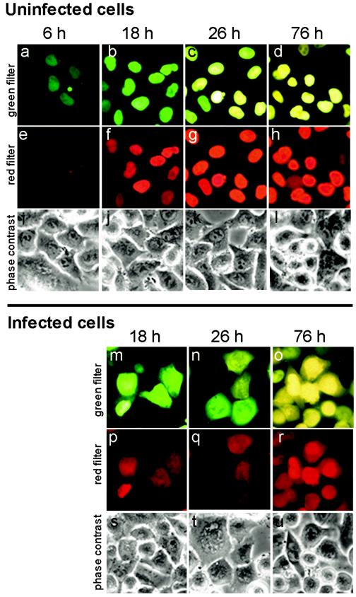
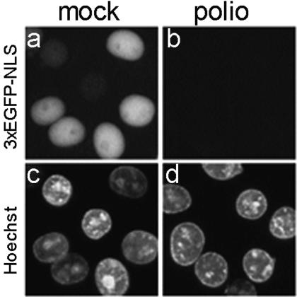
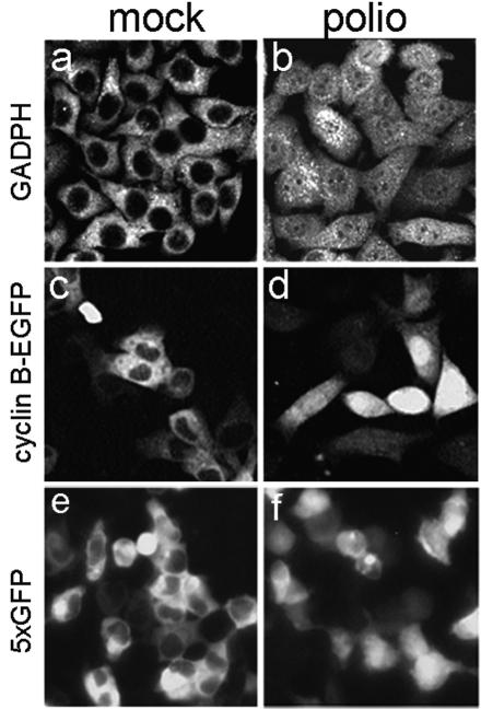
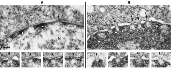
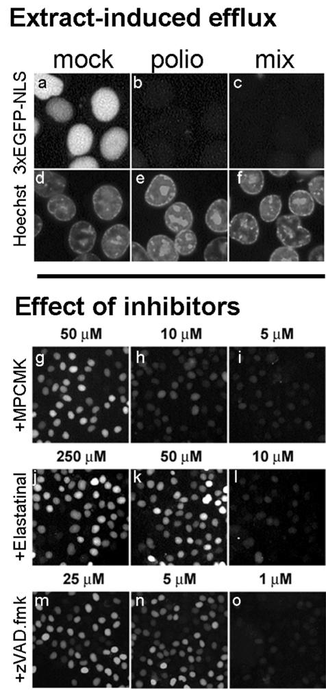
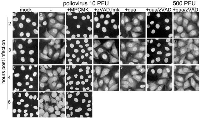
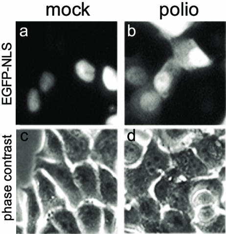
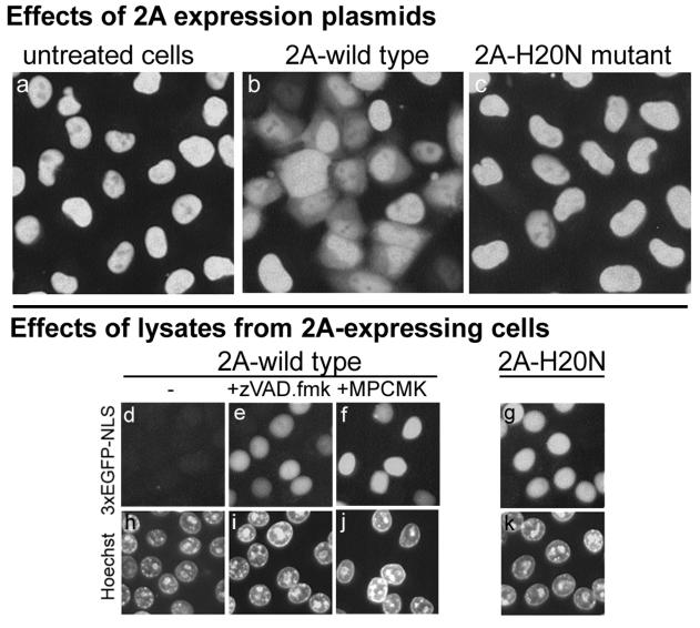
Poliovirus and some other picornaviruses trigger relocation of certain nuclear proteins into the cytoplasm. Here, by using a protein changing its fluorescence color with time and containing a nuclear localization signal (NLS), we demonstrate that the poliovirus-triggered relocation is largely due to the exit of presynthesized nuclear protein into the cytoplasm. The leakiness of the nuclear envelope was also documented by the inability of nuclei from digitonin-permeabilized, virus-infected (but not mock-infected) cells to retain an NLS-containing derivative of green fluorescent protein (GFP). The cytoplasm-to-nucleus traffic was also facilitated during infection, as evidenced by experiments with GAPDH (glyceraldehyde-3-phosphate dehydrogenase), cyclin B1, and an NLS-lacking derivative of GFP, which are predominantly cytoplasmic in uninfected cells. Electron microscopy demonstrated that a bar-like barrier structure in the channel of the nuclear pores, seen in uninfected cells, was missing in the infected cells, giving the impression of fully open pores. Transient expression of poliovirus 2A protease also resulted in relocation of the nuclear proteins. Lysates from poliovirus-infected or 2A-expressing cells induced efflux of 3xEGFP-NLS from the nuclei of permeabilized uninfected cells. This activity was inhibited by the elastase inhibitors elastatinal and N-(methoxysuccinyl)-L-alanyl-L-alanyl-L-prolyl-L-valine chloromethylketone (drugs known also to be inhibitors of poliovirus protease 2A), a caspase inhibitor zVAD(OMe), fmk, and some other protease inhibitors. These data suggest that 2A elicited nuclear efflux, possibly in cooperation with a zVAD(OMe).fmk-sensitive protease. However, poliovirus infection facilitated nuclear protein efflux also in cells deficient in caspase-3 and caspase-9, suggesting that the efflux may occur without the involvement of these enzymes. The biological relevance of nucleocytoplasmic traffic alterations in infected cells is discussed.


Figures

FIG.1.
FIG.1.
Cytoplasmic fluorescence in poliovirus-infected HeLa cells expressing the Timer-NA-NLS fusion was due to the efflux of the presynthesized nuclear protein. In control uninfected Timer-transfected cells (a to l), the newly synthesized protein was accumulated in the nuclei and emitted fluorescence, which shifted with time from green to yellow when inspected with the green filter (a to d) and gradually became visible and increased in intensity when inspected with the red filter (e to h). In poliovirus-infected cells at 4 h p.i. (m to u), fluorescence could be seen not only in the nuclei but also in the cytoplasm, and the colors of nuclear and cytoplasmic fluorescence in a given cell coincided. The exposures were 10, 5, and 2 s for the pictures taken at 6, 18 to 26, and 76 h posttransfection, respectively, due to a marked difference in the fluorescence levels.
FIG. 2.
FIG. 2.
Loss of 3×GFP-NLS from nuclei of permeabilized poliovirus-infected but not mock-infected HeLa-3E cells. At 2 h p.i. (MOI of ∼100 PFU/cell), the monolayers of HeLa-3E cells were treated with digitonin, which by itself permeabilizes the plasma membrane but leaves the nuclear membrane intact, stained with Hoechst 33258, and inspected under fluorescence microscope by using the green (for 3×EGFP-NLS) and blue (for Hoechst) filter cubes. Note that this kind of Hoechst dye is not able to enter the cell through an intact plasma membrane, and therefore it stains only the permeabilized cells.
FIG. 3.
FIG. 3.
Entry of cytoplasmatic proteins in the nucleus after poliovirus infection. Mock-infected (a) and poliovirus-infected (b) Hep-2 cells were stained at 3 h p.i. with monoclonal anti-GAPDH antibodies. The secondary antibodies were Cy3 conjugated. The samples were analyzed with a Zeiss Axiovert 100 confocal microscope. HeLa cells were transfected with a plasmid expressing cyclin B-EGFP and 24 h posttransfection were mock infected (c) or poliovirus infected (d) and inspected under epifluorescence microscope at 2 h p.i. 293 cells were transiently transfected with a plasmid expressing five in-frame copies of EGFP and at 24 h posttransfection were infected with poliovirus. The mock-infected (e) and virus-infected (f) cells were investigated under a fluorescence microscope at 2 h p.i.
FIG. 4.
FIG. 4.
Electron microscopy of nuclear envelope and nuclear pores of uninfected and infected HeLa cells. (A) Uninfected cells. The upper panel shows a segment from a nucleus (N) with nuclear pores (arrows) and small areas of condensed chromatin (asterisks) along the inner nuclear membrane. The sites of the nuclear pore baskets are devoid of condensed chromatin. The lower series of pictures represent examples of nuclear pores, all showing an obstructing bar-like structure in the central channel (arrows). (B) Poliovirus-infected cells at 5 h p.i. The upper panel shows a segment of a nucleus (N) with nuclear pores (arrows), dilated perinuclear space, and heavily condensed chromatin (asterisks). The lower series of pictures show examples of seemingly open nuclear pores (arrows) and the absence of the bar-like closures in the central channel. As in uninfected cells, the condensed chromatin is discontinuous behind the nuclear pores. All pictures in this figure are at an equal magnification. Bar, 200 nm.
FIG. 5.
FIG. 5.
Stimulation of 3×GFP-NLS efflux from nuclei of permeabilized uninfected cells by extracts from poliovirus-infected cells. Monolayers of HeLa-3E cells were permeabilized with digitonin and incubated with 10-fold-diluted S100 extracts from mock-infected (a and d) or poliovirus-infected (b and e) HeLa cells for 1 h at 37°C. In panels c and f, the permeabilized cells were incubated with a 10-fold-diluted mixture of 1 and 9 μl of lysates from infected and mock-infected cells, respectively (the protein concentrations in all probes being equal). The effects of protease inhibitors (g to o) were assayed on permeabilized uninfected HeLa-3E cells by using the extract prepared as described for panels c and f. The extracts were preincubated with the inhibitors for 20 min at 4°C and then applied to the cells for 1 h at 37°C.
FIG. 6.
FIG. 6.
Effects of inhibitors on the infection-triggered protein efflux from the nuclei. HeLa-3E cells were infected as described in Materials and Methods at MOIs of 10 or 500 PFU/cell. The inhibitors were used at the following concentrations: MPCMK, 1 mM; zVAD.fmk, 100 μM; and guanidine-HCl, 2 mM.
FIG. 7.
FIG. 7.
Efflux of EGFP-NLS from the nuclei of poliovirus-infected cells lacking caspase-3 and -9. MCF-Cas9DN cells were transiently transfected with pEGFP-NLS; at 48 h posttransfection they were mock infected (a) or infected with poliovirus at an input MOI of ∼500 PFU/cell (b) and inspected under a fluorescence microscope at 2 h p.i.
FIG. 8.
FIG. 8.
Effect of poliovirus 2A protease on the relocation of 3×GFP-NLS from the nuclei of uninfected cells. In the upper set of panels (a to c), HeLa-3E cells were untreated (a) or transfected with plasmids expressing either the wild-type poliovirus 2A (b) or 2A harboring a protease inactivating H20N mutation (c). The pictures were taken 12 h after the transfection. In the lower set of panels (d to k), twofold-diluted S15 extracts from HeLa cells expressing wild-type (d to f and h to j) and mutant (g and k) 2A were applied to the permeabilized uninfected HeLa-3E cells for 1 h at 37°C. When used, zVAD.fmk and MPCMK were at 5 and 50 μM, respectively, and preincubated with the extracts for 20 min at 4°C.

References

    1. Adam, S. A., S. R. Marr, and L. Gerace. 1990. Nuclear protein import in permeabilized mammalian cells requires soluble cytoplasmic factors. J. Cell Biol. 111:807-816. - PMC - PubMed
    1. Agol, V. I., G. A. Belov, K. Bienz, D. Egger, M. S. Kolesnikova, N. T., Raikhlin, L. I. Romanova, E. A. Smirnova, and E. A. Tolskaya. 1998. Two types of death of poliovirus-infected cells: caspase involvement in the apoptosis but not cytopathic effect. Virology 252:342-353. - PubMed
    1. Aminev, A. G., S. P. Amineva, and A. C. Palmenberg. 2003. Encephalomyocarditis viral protein 2A localizes to nucleoli and inhibits cap-dependent mRNA translation. Virus Res. 95:45-57. - PubMed
    1. Aminev, A. G., S. P. Amineva, and A. C. Palmenberg. 2003. Encephalomyocarditis virus (EMCV) proteins 2A and 3BCD localize to nuclei and inhibit cellular mRNA transcription but not rRNA transcription. Virus Res. 95:59-73. - PubMed
    1. Barton, D. J., and J. B. Flanegan. 1993. Coupled translation and replication of poliovirus RNA in vitro: synthesis of functional 3D polymerase and infectious virus. J. Virol. 67:822-831. - PMC - PubMed

Publication types

MeSH terms