. 2004 Sep;5(9):906-13.
doi: 10.1038/sj.embor.7400240.

Position of the CrPV IRES on the 40S subunit and factor dependence of IRES/80S ribosome assembly

Affiliations

Position of the CrPV IRES on the 40S subunit and factor dependence of IRES/80S ribosome assembly

Tatyana V Pestova et al. EMBO Rep. 2004 Sep.

Abstract

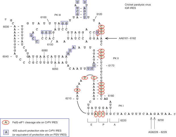
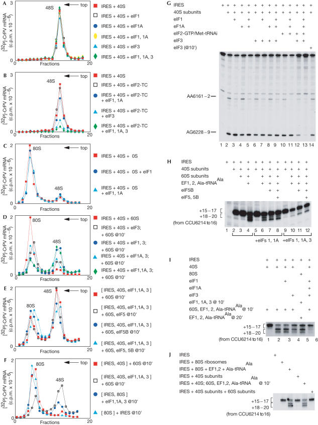
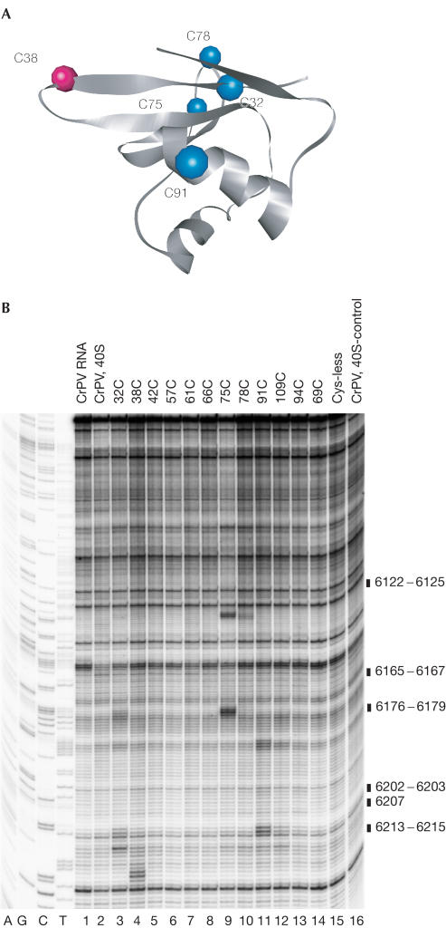
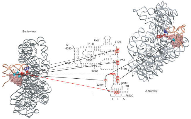
The cricket paralysis virus intergenic region internal ribosomal entry site (CrPV IGR IRES) can assemble translation initiation complexes by binding to 40S subunits without Met-tRNA(Met)(i) and initiation factors (eIFs) and then by joining directly with 60S subunits, yielding elongation-competent 80S ribosomes. Here, we report that eIF1, eIF1A and eIF3 do not significantly influence IRES/40S subunit binding but strongly inhibit subunit joining and the first elongation cycle. The IRES can avoid their inhibitory effect by its ability to bind directly to 80S ribosomes. The IRES's ability to bind to 40S subunits simultaneously with eIF1 allowed us to use directed hydroxyl radical cleavage to map its position relative to the known position of eIF1. A connecting loop in the IRES's pseudoknot (PK) III domain, part of PK II and the entire domain containing PK I are solvent-exposed and occupy the E site and regions of the P site that are usually occupied by Met-tRNA(Met)(i).


Figures

Figure 1
Figure 1
Structure of the CrPV IGR IRES (Kanamori & Nakashima, 2001; Jan & Sarnow, 2002) showing toe-prints (black arrows) due to binding of 40S subunits and 80S ribosomes (Wilson et al, 2000) and sites of cleavage (red circles) induced by 40Ssubunit-bound Fe(II)-eIF1 derivatives (this work). Sites protected by 40S subunits from chemical and enzymatic cleavage (blue squares) in CrPV and PSIV IRESs (Jan & Sarnow, 2002; Nishiyama et al, 2003) have been combined.
Figure 2
Figure 2
Influence of initiation factors on ribosomal binding to the IRES. Sucrose density gradient analysis of the influence of eIFs (as indicated) on (A,B) IRES–40S subunit binding and (C–F) joining of 60S subunits to IRES–40S–eIF complexes. The positions of 48S and 80S complexes are indicated. Fractions from the upper parts of gradients have been omitted for clarity. (G) Toe-print analysis of 40S subunit binding to IRES RNA in the presence of eIFs as indicated. The 40S-dependent toe-prints are at AA6161–2 and AG6228–9. (H–J) Toe-print analysis of ribosome translocation on the IRES in reactions containing components as indicated. The toe-prints seen on binding of 40S subunits or 80S ribosomes and following ribosomal translocation are indicated to the left.
Figure 3
Figure 3
Formation of eIF1–40S subunit–IRES complexes. (A) Binding of 40S subunits and the IRES to T7-Tag antibody agarose-immobilized eIF1. Ribosomal protein S6, eIF1 and the IRES were visualized by western blotting, Coomassie staining and primer extension, respectively. (BD) Primer extension analysis of directed hydroxyl radical cleavage of 18S rRNA helices 23, 24 and 44, respectively, in 40S–eIF1–IRES complexes from Fe(II) tethered to positions on the surface of eIF1, as indicated. Reaction mixtures marked ‘40S, IRES' did not contain eIF1, those marked ‘40S' lacked eIF1 and the IRES, and those marked ‘Cys-less' contained the cysteine-less eIF1 mutant. Lanes A, G, C and T depict 18S rRNA sequence obtained using the same primer. The positions of cleaved nucleotides are indicated to the right of each panel.
Figure 4
Figure 4
Directed hydroxyl radical cleavage of the CrPV IRES bound to 40S subunits. (A) Ribbon diagram of the structured domain of human eIF1. Coloured spheres indicate positions of cysteines on the surface of eIF1 from which hydroxyl radicals cleaved the IRES. (B) Primer extension analysis of directed hydroxyl radical cleavage of the IRES in eIF1–40S–IRES complexes from Fe(II) tethered to surface positions on eIF1, as indicated (lanes 3–14). Control reactions were carried out without eIF1 (lane 16) or with cysteine-less eIF1 mutant (lane 15). Hydroxyl radical cleavage was not induced in reactions shown in lanes 1 and 2. The positions of cleaved nucleotides are indicated to the right of the panel.
Figure 5
Figure 5
Orientation of the CrPV IRES bound to a 40S subunit relative to eIF1. E- and Asite views of the position of eIF1 (pink) on the small ribosomal subunit (grey) relative to initiator tRNA (coral) and A- and P-site mRNA (red) were modelled as described by Lomakin et al (2003). eIF1 residues from which hydroxyl radicals cleaved the IRES are shown in turquoise (32, 78, 75 and 91) and red (38). The anticodon bases of Psite tRNA are dark blue. The strong sites of hydroxyl radical cleavages in the IRES (red circles) are connected by solid lines to the positions on eIF1 from which they occurred. Weaker cleavage sites are connected by dashed lines.

References

    1. Carter AP et al. (2001) Crystal structure of an initiation factor bound to the 30S ribosomal subunit. Science 291: 498–501 - PubMed
    1. Fletcher CM, Pestova TV, Hellen CUT, Wagner G (1999) Structure and interactions of the translation initiation factor eIF1. EMBO J 18: 2631–2637 - PMC - PubMed
    1. Jan E, Sarnow P (2002) Factorless ribosome assembly on the internal ribosome entry site of cricket paralysis virus. J Mol Biol 324: 889–902 - PubMed
    1. Kanamori Y, Nakashima N (2001) A tertiary structure model of the internal ribosome entry site (IRES) for methionine-independent initiation of translation. RNA 7: 266–274 - PMC - PubMed
    1. Lomakin IB, Kolupaeva VG, Marintchev A, Wagner G, Pestova TV (2003) Position of eukaryotic initiation factor eIF1 on the 40S ribosomal subunit determined by directed hydroxyl radical probing. Genes Dev 17: 2786–2797 - PMC - PubMed

Publication types