

Melanopsin--shedding light on the elusive circadian photopigment
- PMID: 15332341
- PMCID: PMC2376768
- DOI: 10.1081/cbi-120037816
Melanopsin--shedding light on the elusive circadian photopigment
Abstract
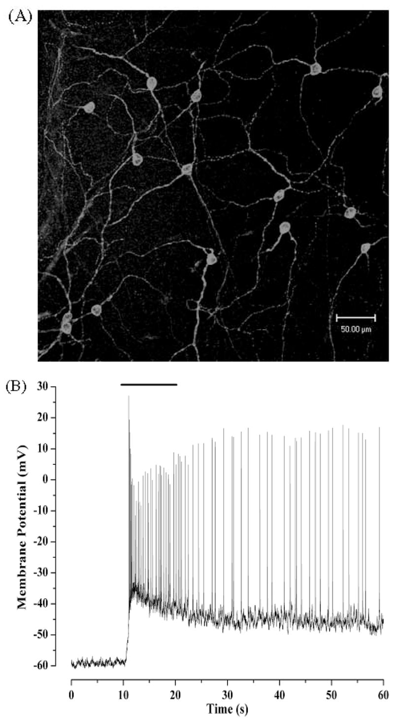
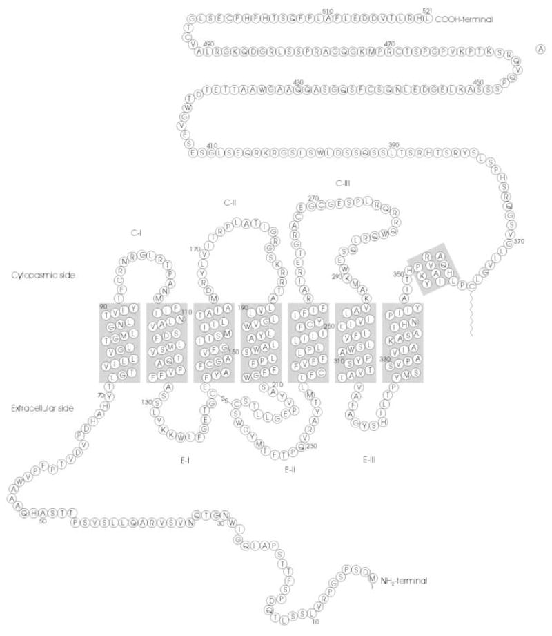
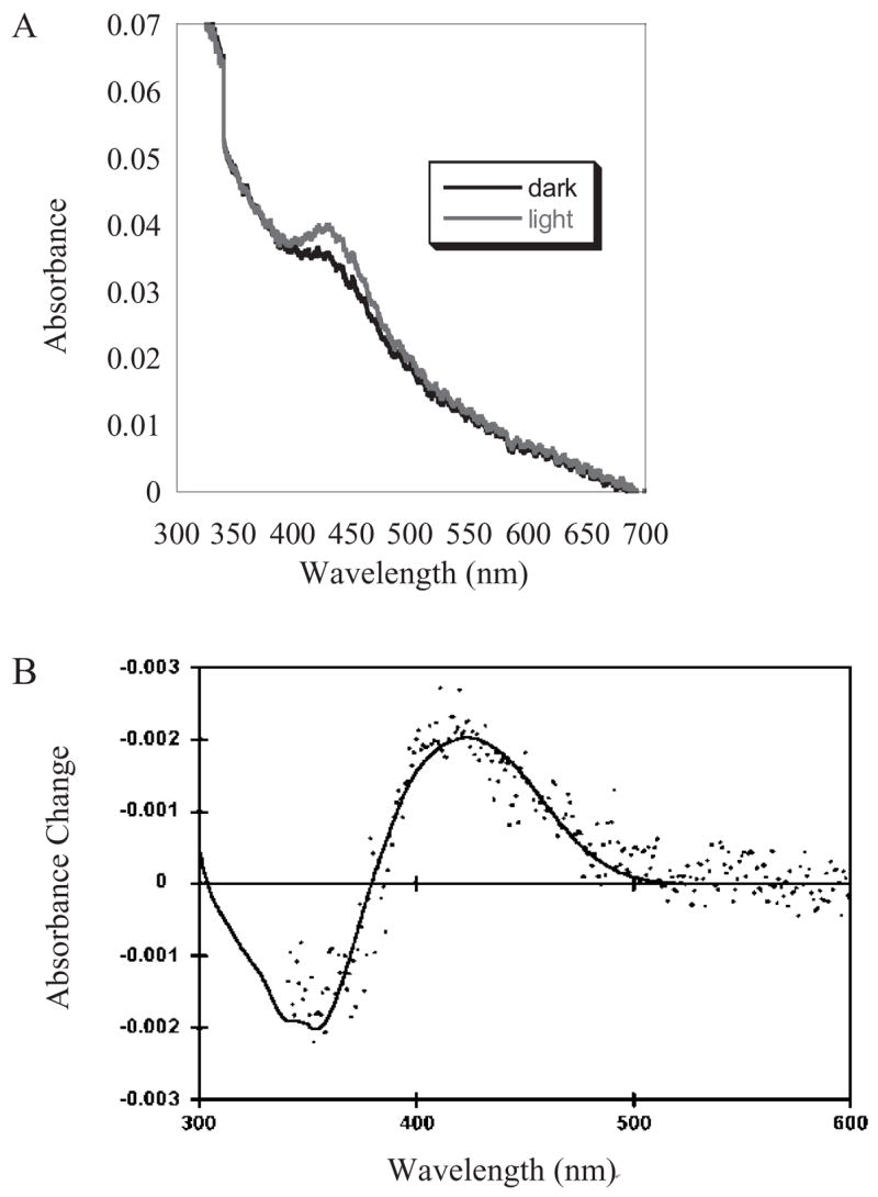
Circadian photoentrainment is the process by which the brain's internal clock becomes synchronized with the daily external cycle of light and dark. In mammals, this process is mediated exclusively by a novel class of retinal ganglion cells that send axonal projections to the suprachiasmatic nuclei (SCN), the region of the brain that houses the circadian pacemaker. In contrast to their counterparts that mediate image-forming vision, SCN-projecting RGCs are intrinsically sensitive to light, independent of synaptic input from rod and cone photoreceptors. The recent discovery of these photosensitive RGCs has challenged the long-standing dogma of retinal physiology that rod and cone photoreceptors are the only retinal cells that respond directly to light and has explained the perplexing finding that mice lacking rod and cone photoreceptors can still reliably entrain their circadian rhythms to light. These SCN-projecting RGCs selectively express melanopsin, a novel opsin-like protein that has been proposed as a likely candidate for the photopigment in these cells. Research in the past three years has revealed that disruption of the melanopsin gene impairs circadian photo- entrainment, as well as other nonvisual responses to light such as the pupillary light reflex. Until recently, however, there was no direct demonstration that melanopsin formed a functional photopigment capable of catalyzing G-protein activation in a light-dependent manner. Our laboratory has recently succeeded in expressing melanopsin in a heterologous tissue culture system and reconstituting a pigment with the 11-cis-retinal chromophore. In a reconstituted biochemical system, the reconstituted melanopsin was capable of activating transducin, the G-protein of rod photoreceptors, in a light-dependent manner. The absorbance spectrum of this heterologously expressed melanopsin, however, does not match that predicted by previous behavioral and electophysiological studies. Although melanopsin is clearly the leading candidate for the elusive photopigment of the circadian system, further research is needed to resolve the mystery posed by its absorbance spectrum and to fully elucidate its role in circadian photoentrainment.
Figures

Similar articles
-
Melanopsin forms a functional short-wavelength photopigment.Biochemistry. 2003 Nov 11;42(44):12734-8. doi: 10.1021/bi035418z. Biochemistry. 2003. PMID: 14596587
-
Synaptic inputs to retinal ganglion cells that set the circadian clock.Eur J Neurosci. 2006 Aug;24(4):1117-23. doi: 10.1111/j.1460-9568.2006.04999.x. Eur J Neurosci. 2006. PMID: 16930437 Free PMC article.
-
Melanopsin-containing retinal ganglion cells: architecture, projections, and intrinsic photosensitivity.Science. 2002 Feb 8;295(5557):1065-70. doi: 10.1126/science.1069609. Science. 2002. PMID: 11834834 Free PMC article.
-
Melanopsin: a novel photopigment involved in the photoentrainment of the brain's biological clock?Ann Med. 2002;34(5):401-7. doi: 10.1080/078538902320772151. Ann Med. 2002. PMID: 12452484 Review.
-
Roles of PACAP-containing retinal ganglion cells in circadian timing.Int Rev Cytol. 2006;251:1-39. doi: 10.1016/S0074-7696(06)51001-0. Int Rev Cytol. 2006. PMID: 16939776 Review.
Cited by
-
Phototransduction in ganglion-cell photoreceptors.Pflugers Arch. 2007 Aug;454(5):849-55. doi: 10.1007/s00424-007-0242-2. Epub 2007 Mar 10. Pflugers Arch. 2007. PMID: 17351786 Review.
-
In synch but not in step: Circadian clock circuits regulating plasticity in daily rhythms.Neuroscience. 2016 Apr 21;320:259-80. doi: 10.1016/j.neuroscience.2016.01.072. Epub 2016 Feb 6. Neuroscience. 2016. PMID: 26861419 Free PMC article. Review.
-
Deep Diversity: Extensive Variation in the Components of Complex Visual Systems across Animals.Cells. 2022 Dec 8;11(24):3966. doi: 10.3390/cells11243966. Cells. 2022. PMID: 36552730 Free PMC article. Review.
-
Biological clocks: their relevance to immune-allergic diseases.Clin Mol Allergy. 2018 Jan 10;16:1. doi: 10.1186/s12948-018-0080-0. eCollection 2018. Clin Mol Allergy. 2018. PMID: 29344005 Free PMC article. Review.
-
Perspectives of colon-specific drug delivery in the management of morning symptoms of rheumatoid arthritis.Inflammopharmacology. 2023 Feb;31(1):253-264. doi: 10.1007/s10787-022-01120-w. Epub 2022 Dec 21. Inflammopharmacology. 2023. PMID: 36544060 Review.
References
-
- Belenky MA, Smeraski CA, Provencio I, Sollars PJ, Pickard GE. Melanopsin retinal ganglion cells receive bipolar and amacrine cell synapses. Journal of Comparative Neurology. 2003;460:380–393. - PubMed
-
- Bellingham J, Foster RG. Opsins and mammalian photoentrainment. Cell and Tissue Research. 2002;309:57–71. - PubMed
-
- Berson DM, Dunn FA, Takao M. Phototransduction by retinal ganglion cells that set the circadian clock. Science. 2002;295:1070–1073. - PubMed
-
- Ceriani MF, Darlington TK, Staknis D, Mas P, Petti AA, Weitz CJ, Kay SA. Light-dependent sequestration of TIMELESS by CRYPTOCHROME. Science. 1999;285:553–556. - PubMed
-
- Chen P, Hao W, Rife L, Wang XP, Shen D, Chen J, Ogden T, Van Boemel GB, Wu L, Yang M, Fong HK. A photic visual cycle of rhodopsin regeneration is dependent on Rgr. Nauret Geneicst. 2001;28:256–260. - PubMed
Publication types
MeSH terms
Substances
Grants and funding
LinkOut - more resources
Full Text Sources
Other Literature Sources
Miscellaneous