

Morules in endometrial carcinoma and benign endometrial lesions differ from squamous differentiation tissue and are not infected with human papillomavirus
- PMID: 15333650
- PMCID: PMC1770414
- DOI: 10.1136/jcp.2004.017996
Morules in endometrial carcinoma and benign endometrial lesions differ from squamous differentiation tissue and are not infected with human papillomavirus
Abstract
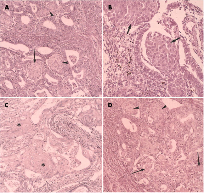
Background: Squamous differentiation/squamous metaplasia is often associated with endometrial adenocarcinoma and benign lesions, such as endometrial hyperplasia and chronic endometritis. Morules have distinct histological characteristics, and are referred to as squamous metaplasia or squamoid metaplasia.
Aim: To focus on the histological characteristics of morules and clarify the difference between morules and squamous differentiation.
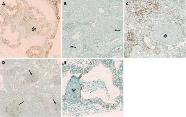
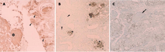
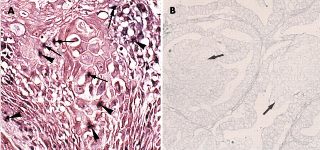
Materials/methods: Twenty endometrioid carcinomas with morules or squamous differentiation, five adenosquamous carcinomas, and eight non-carcinomatous endometrial lesions with morules were investigated. Numerous antibodies for epithelial membrane antigen (EMA), involucrin, cytokeratins, neuropeptides, and oncofetal antigens were used for immunohistochemistry. In situ hybridisation and polymerase chain reaction were used to detect human papillomavirus (HPV).
Results: The morules observed were uniform cell clusters, with no squamous differentiation. They were immunonegative for epithelial antigens including involucrin, EMA, and cytokeratins, but were positive for neurone specific enolase. A few morules were immunopositive for acetylcholine esterase, and one case was positive for somatostatin; neither oncofetal nor proliferative cell markers, including blood group A, B, and AB, or other neuropeptides were demonstrated in the morules. HPV DNA was not found in either the morules in the carcinomas or in the benign lesions. However, true squamous differentiation tissue in four endometrioid carcinomas and two adenosquamous carcinomas was HPV positive using in situ hybridisation.
Conclusion: Morules are histologically distinct from squamous metaplasia/squamous differentiation tissue. Morules are thought to be neuroectodermal-like cell clusters, and are not infected with HPV. In contrast, some of the true squamous differentiation tissue was associated with HPV infection.
Figures
Similar articles
-
Morules and morule-like features associated with carcinomas in various organs: report with immunohistochemical and molecular studies.J Clin Pathol. 2006 Jan;59(1):95-100. doi: 10.1136/jcp.2005.026237. J Clin Pathol. 2006. PMID: 16394288 Free PMC article.
-
Human papillomavirus and mixed epithelial tumors of the endometrium.Hum Pathol. 1998 Apr;29(4):383-9. doi: 10.1016/s0046-8177(98)90120-4. Hum Pathol. 1998. PMID: 9563789
-
Squamous metaplasia of the endometrium associated with HPV 6 and 11.Gynecol Oncol. 1997 Jul;66(1):141-5. doi: 10.1006/gyno.1997.4731. Gynecol Oncol. 1997. PMID: 9234935
-
Histopathology of functional and neoplastic changes in cervix and endometrium.Verh Dtsch Ges Pathol. 1997;81:240-4. Verh Dtsch Ges Pathol. 1997. PMID: 9474876 Review.
-
Carcinoma of the lung in Okinawa, Japan: with special reference to squamous cell carcinoma and squamous metaplasia.Pathol Int. 1997 Oct;47(10):659-72. doi: 10.1111/j.1440-1827.1997.tb04439.x. Pathol Int. 1997. PMID: 9361099 Review.
Cited by
-
Morules and morule-like features associated with carcinomas in various organs: report with immunohistochemical and molecular studies.J Clin Pathol. 2006 Jan;59(1):95-100. doi: 10.1136/jcp.2005.026237. J Clin Pathol. 2006. PMID: 16394288 Free PMC article.
-
Human papillomavirus in endometrial adenocarcinomas: infectious agent or a mere "passenger"?Infect Dis Obstet Gynecol. 2007;2007:60549. doi: 10.1155/2007/60549. Infect Dis Obstet Gynecol. 2007. PMID: 18274613 Free PMC article.
-
Does endometrial morular metaplasia represent odontogenic differentiation?Virchows Arch. 2021 Sep;479(3):607-616. doi: 10.1007/s00428-021-03060-2. Epub 2021 Mar 5. Virchows Arch. 2021. PMID: 33666744 Free PMC article.
-
Association between human papillomavirus and endometrial adenocarcinoma.Med Oncol. 2013;30(3):597. doi: 10.1007/s12032-013-0597-5. Epub 2013 Jun 25. Med Oncol. 2013. PMID: 23797769
-
Possible Risk Factors of Pulmonary Metastases in Patients With International Federation of Gynecology and Obstetrics Stage I Endometrioid-Type Endometrial Cancer.Int J Gynecol Cancer. 2017 Jul;27(6):1206-1215. doi: 10.1097/IGC.0000000000001002. Int J Gynecol Cancer. 2017. PMID: 28448305 Free PMC article.
References
-
- Anderson MC, Robby SJ, Russell P, et al. Endometritis, metaplasia, polyp and miscellaneous changes. In: Robby SJ, Anderson MC, Russell P, eds. Pathology of the female reproductive tract. London: Churchill Livingstone, 2002:285–303.
-
- Buckley CH. Normal endometrium and non-proliferative conditions of the endometrium. In: Fox H, Wells M, eds. Haines and Taylor obstetrical and gynaecological pathology. 5th ed. London: Churchill Livingstone, 2003:391–441.
-
- Novak ER, Woodruff JD. Squamous metaplasia or epidermization. In: Novak ER, Woodruff JD, eds. Novak’s gynecologic and obstetric pathology with clinical and endocrine relations. 8th ed. Philadelphia: WB Saunders, 1979:199–200.
-
- Prat J. Female reproductive system. In: Damjanov I, Linder J, eds. Anderson’s pathology. 10th ed. St Louis: Mosby, 1996:2231–309.
-
- Silverberg SG, Kurman RJ. Endometrial carcinoma. In: Silverberg SG, Kurman RJ, eds. Atlas of tumor pathology, 3rd Series, Fascicles 3. Tumors of the uterine corpus and gestational trophoblastic disease. Washington, DC: Armed Forces Institute of Pathology, 1991:47–89.
MeSH terms
Substances
LinkOut - more resources
Full Text Sources
Research Materials
Miscellaneous