. 2004 Sep 7;101(36):13163-7.
doi: 10.1073/pnas.0403045101. Epub 2004 Aug 27.

Incorporation of a single His residue by rational design enables thiol-ester hydrolysis by human glutathione transferase A1-1

Affiliations

Incorporation of a single His residue by rational design enables thiol-ester hydrolysis by human glutathione transferase A1-1

Sofia Hederos et al. Proc Natl Acad Sci U S A. .

Abstract

A strategy for rational enzyme design is reported and illustrated by the engineering of a protein catalyst for thiol-ester hydrolysis. Five mutants of human glutathione (GSH; gamma-Glu-Cys-Gly) transferase A1-1 were designed in the search for a catalyst and to provide a set of proteins from which the reaction mechanism could be elucidated. The single mutant A216H catalyzed the hydrolysis of the S-benzoyl ester of GSH under turnover conditions with a k(cat)/K(M) of 156 M(-1) x min(-1), and a catalytic proficiency of >10(7) M(-1) when compared with the first-order rate constant of the uncatalyzed reaction. The wild-type enzyme did not hydrolyze the substrate, and thus, the introduction of a single histidine residue transformed the wild-type enzyme into a turnover system for thiol-ester hydrolysis. By kinetic analysis of single, double, and triple mutants, as well as from studies of reaction products, it was established that the enzyme A216H catalyzes the hydrolysis of the thiol-ester substrate by a mechanism that includes an acyl intermediate at the side chain of Y9. Kinetic measurements and the crystal structure of the A216H GSH complex provided compelling evidence that H216 acts as a general-base catalyst. The introduction of a single His residue into human GSH transferase A1-1 created an unprecedented enzymatic function, suggesting a strategy that may be of broad applicability in the design of new enzymes. The protein catalyst has the hallmarks of a native enzyme and is expected to catalyze various hydrolytic, as well as transesterification, reactions.


Figures

Fig. 1.
Fig. 1.
Detail of the crystal structure (23) of the complex between S-benzylglutathione and GST A1-1 (PDB ID code 1GUH) highlighting side-chain positions of residues exploited in the design of the catalytic machinery for thiol-ester hydrolysis. Based on this structure, Y9, A216, and F220 were expected to be in close proximity to the thiol-ester functionality of the putative substrate GSB. The figure was created by using pymol (36).
Fig. 2.
Fig. 2.
Saturation kinetic signature of A216H-catalyzed hydrolysis of GSB and HPLC traces recorded under reaction conditions demonstrating benzoic acid production in the A216H-catalyzed reaction but not in the presence of wild-type GST A1-1 (Inset; B, benzoic acid). The kinetic studies were made at a concentration of 5 μM A216H in 100 mM sodium phosphate at 298 K and pH 7. Rates were determined by spectroscopic monitoring of the thiol-ester band of GSB at 266 nm (ε266 = 7,889 M–1·cm–1).
Fig. 3.
Fig. 3.
Plot of log v versus pH of the A216H-catalyzed hydrolysis of GSB, suggesting that catalysis depends on the unprotonated form of a catalytic residue with a pKa of ≈6. The observed deviation from linearity at high pH remains to be understood but is not found in the wild-type enzyme, and thus, is linked to the new function of A216H. The experimental concentrations were 5 μM A216H, 75 μM GSB in 100 mM sodium acetate, or 100 mM sodium phosphate at 298 K.
Fig. 4.
Fig. 4.
Trapping of the acyl intermediate by MeOH increases the reaction rate and leads to the production of methylbenzoate (shown by a HPLC trace; B, benzoic acid; MeB, methylbenzoate). We used 5 μM A216H and 75 μM GSB in 100 mM sodium phosphate buffer at pH 7 and 298 K.
Fig. 5.
Fig. 5.
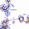
Detail of the crystal structure of the complex between S-benzylglutathione and A216H, showing the distance between the phenolic oxygen of Y9 and the side chain of H216 to be ≈7 Å, which is too long for nucleophilic catalysis. The distance between the phenolic oxygen of Y9 and the sulfur atom of S-benzylglutathione was 3 Å, in good agreement with a model in which Y9 attacks the carbonyl carbon of GSB in the second step of the reaction (details not shown). The protein was cocrystallized with S-benzylglutathione, but in the crystal structure no density for the benzyl group is visible. The figure was created by using the programs o (37) and molray (38).

Similar articles

Cited by

References

    1. Cedrone, F., Menez, A. & Quemeneur, E. (2000) Curr. Opin. Struct. Biol. 10, 405–410. - PubMed
    1. Penning, T. M. & Jez, J. M. (2001) Chem. Rev. 101, 3027–3046. - PubMed
    1. Qi, D. F., Tann, C. M., Haring, D. & Distefano, M. D. (2001) Chem. Rev. 101, 3081–3111. - PubMed
    1. Jez, J. M. & Penning, T. M. (1998) Biochemistry 37, 9695–9703. - PubMed
    1. Nilsson, L. O., Gustafsson, A. & Mannervik, B. (2000) Proc. Natl. Acad. Sci. USA 97, 9408–9412. - PMC - PubMed

Publication types

Associated data

LinkOut - more resources