

System for prostate brachytherapy and biopsy in a standard 1.5 T MRI scanner
- PMID: 15334592
- PMCID: PMC2396258
- DOI: 10.1002/mrm.20138
System for prostate brachytherapy and biopsy in a standard 1.5 T MRI scanner
Abstract
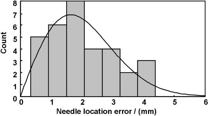
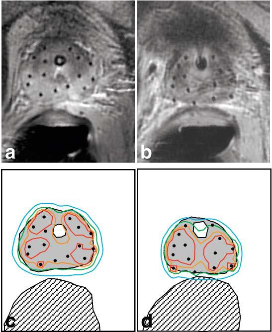
A technique for transperineal high-dose-rate (HDR) prostate brachytherapy and needle biopsy in a standard 1.5 T MRI scanner is demonstrated. In each of eight procedures (in four patients with intermediate to high risk localized prostate cancer), four MRI-guided transperineal prostate biopsies were obtained followed by placement of 14-15 hollow transperineal catheters for HDR brachytherapy. Mean needle-placement accuracy was 2.1 mm, 95% of needle-placement errors were less than 4.0 mm, and the maximum needle-placement error was 4.4 mm. In addition to guiding the placement of biopsy needles and brachytherapy catheters, MR images were also used for brachytherapy treatment planning and optimization. Because 1.5 T MR images are directly acquired during the interventional procedure, dependence on deformable registration is reduced and online image quality is maximized.
Copyright 2004 Wiley-Liss, Inc.
Figures

Similar articles
-
Open MR-guided high-dose-rate (HDR) prostate brachytherapy: feasibility and initial experiences open MR-guided high-dose-rate (HDR) prostate brachytherapy.Pathol Oncol Res. 2011 Jun;17(2):315-24. doi: 10.1007/s12253-010-9319-x. Epub 2011 Jan 11. Pathol Oncol Res. 2011. PMID: 21221879 Clinical Trial.
-
Robotic MR-guided high dose rate brachytherapy needle implantation in the prostate (ROBiNSon)-a proof-of-concept study.Phys Med Biol. 2024 Aug 21;69(17). doi: 10.1088/1361-6560/ad69f8. Phys Med Biol. 2024. PMID: 39084657
-
MRI targeted single fraction HDR Brachytherapy for localized Prostate Carcinoma: a feasibility study of focal radiation therapy (ProFocAL).Eur Radiol. 2020 Apr;30(4):2072-2081. doi: 10.1007/s00330-019-06505-0. Epub 2019 Dec 11. Eur Radiol. 2020. PMID: 31828412
-
MR-guided interventions for prostate cancer.Magn Reson Imaging Clin N Am. 2005 Aug;13(3):491-504. doi: 10.1016/j.mric.2005.04.012. Magn Reson Imaging Clin N Am. 2005. PMID: 16084415 Review.
-
Clinical evaluation of an MRI-to-ultrasound deformable image registration algorithm for prostate brachytherapy.Brachytherapy. 2019 Jan-Feb;18(1):95-102. doi: 10.1016/j.brachy.2018.08.006. Epub 2018 Oct 2. Brachytherapy. 2019. PMID: 30287271 Review.
Cited by
-
Towards Safe In Situ Needle Manipulation for Robot Assisted Lumbar Injection in Interventional MRI.Rep U S. 2021 Sep-Oct;2021:1835-1842. doi: 10.1109/iros51168.2021.9636220. Epub 2021 Dec 16. Rep U S. 2021. PMID: 35173994 Free PMC article.
-
GantryMate: A Modular MR-Compatible Assistance System for MR-Guided Needle Interventions.Tomography. 2019 Jun;5(2):266-273. doi: 10.18383/j.tom.2019.00007. Tomography. 2019. PMID: 31245548 Free PMC article.
-
Deformable registration of trans-rectal ultrasound (TRUS) and magnetic resonance imaging (MRI) for focal prostate brachytherapy.Int J Comput Assist Radiol Surg. 2016 Jun;11(6):1015-23. doi: 10.1007/s11548-016-1380-9. Epub 2016 Mar 26. Int J Comput Assist Radiol Surg. 2016. PMID: 27017500
-
Preclinical evaluation of an MRI-compatible pneumatic robot for angulated needle placement in transperineal prostate interventions.Int J Comput Assist Radiol Surg. 2012 Nov;7(6):949-57. doi: 10.1007/s11548-012-0750-1. Epub 2012 Jun 8. Int J Comput Assist Radiol Surg. 2012. PMID: 22678723 Free PMC article.
-
MR imaging-guided interventions in the genitourinary tract: an evolving concept.Magn Reson Imaging Clin N Am. 2010 Feb;18(1):11-28. doi: 10.1016/j.mric.2009.09.002. Magn Reson Imaging Clin N Am. 2010. PMID: 19962090 Free PMC article.
References
-
- Cancer facts & figures. 2003. American Cancer Society; Atlanta: 2003.
-
- Chodak GW, Thisted RA, Gerber GS, Johansson JE, Adolfsson J, Jones GW, Chisholm GD, Moskovitz B, Livne PM, Warner J. Results of conservative management of clinically localized prostate cancer. N Engl J Med. 1994;330:242–248. - PubMed
-
- Potosky AL, Legler J, Albertsen PC, Stanford JL, Gilliland FD, Hamilton AS, Eley JW, Stephenson RA, Harlan LC. Health outcomes after prostatectomy or radiotherapy for prostate cancer: results from the Prostate Cancer Outcomes Study. J Natl Cancer Inst. 2000;92:1582–1592. - PubMed
-
- Yu KK, Hricak H. Imaging prostate cancer. Radiol Clin North Am. 2000;38:59–85. - PubMed
Publication types
MeSH terms
Grants and funding
LinkOut - more resources
Full Text Sources
Other Literature Sources
Medical
