

Middle and inferior temporal gyrus gray matter volume abnormalities in chronic schizophrenia: an MRI study
- PMID: 15337650
- PMCID: PMC2793337
- DOI: 10.1176/appi.ajp.161.9.1603
Middle and inferior temporal gyrus gray matter volume abnormalities in chronic schizophrenia: an MRI study
Abstract
Objective: The middle temporal gyrus and inferior temporal gyrus subserve language and semantic memory processing, visual perception, and multimodal sensory integration. Functional deficits in these cognitive processes have been well documented in patients with schizophrenia. However, there have been few in vivo structural magnetic resonance imaging (MRI) studies of the middle temporal gyrus and inferior temporal gyrus in schizophrenia.
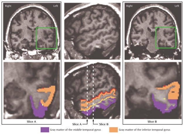
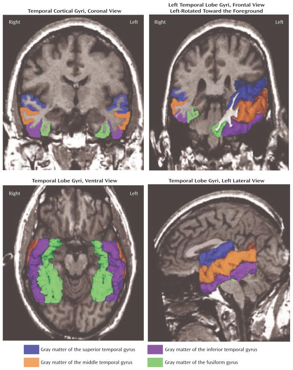
Method: Middle temporal gyrus and inferior temporal gyrus gray matter volumes were measured in 23 male patients diagnosed with chronic schizophrenia and 28 healthy male subjects by using high-spatial-resolution MRI. For comparison, superior temporal gyrus and fusiform gyrus gray matter volumes were also measured. Correlations between these four regions and clinical symptoms were also investigated.
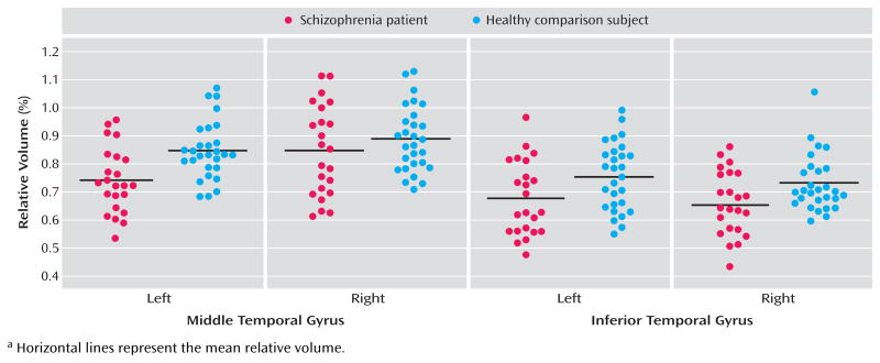
Results: Relative to healthy subjects, the patients with chronic schizophrenia showed gray matter volume reductions in the left middle temporal gyrus (13% difference) and bilateral inferior temporal gyrus (10% difference in both hemispheres). In addition, the patients showed gray matter volume reductions in the left superior temporal gyrus (13% difference) and bilateral fusiform gyrus (10% difference in both hemispheres). More severe hallucinations were significantly correlated with smaller left hemisphere volumes in the superior temporal gyrus and middle temporal gyrus.
Conclusions: These results suggest that patients with schizophrenia evince reduced gray matter volume in the left middle temporal gyrus and bilateral reductions in the inferior temporal gyrus. In conjunction with findings of left superior temporal gyrus reduction and bilateral fusiform gyrus reductions, these data suggest that schizophrenia may be characterized by left hemisphere-selective dorsal pathophysiology and bilateral ventral pathophysiology in temporal lobe gray matter.
Figures
References
-
- Cabeza R, Nyberg L. Imaging cognition, II: an empirical review of 275 PET and fMRI studies. J Cogn Neurosci. 2000;12:1–47. - PubMed
-
- Tranel D, Damasio H, Damasio AR. A neural basis for the retrieval of conceptual knowledge. Neuropsychologia. 1997;35:1329–1339. - PubMed
-
- Chao LL, Haxby JV, Martin A. Attribute-based neural substrates in temporal cortex for perceiving and knowing about objects. Nat Neurosci. 1999;2:913–919. - PubMed
Publication types
MeSH terms
Grants and funding
LinkOut - more resources
Full Text Sources
Medical
