. 2004 Aug 30;166(5):685-96.
doi: 10.1083/jcb.200406181.

Sorting of a nonmuscle tropomyosin to a novel cytoskeletal compartment in skeletal muscle results in muscular dystrophy

Affiliations

Sorting of a nonmuscle tropomyosin to a novel cytoskeletal compartment in skeletal muscle results in muscular dystrophy

Anthony J Kee et al. J Cell Biol. .

Abstract

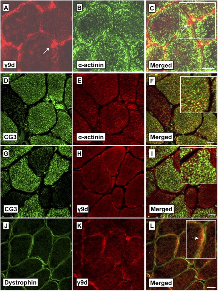
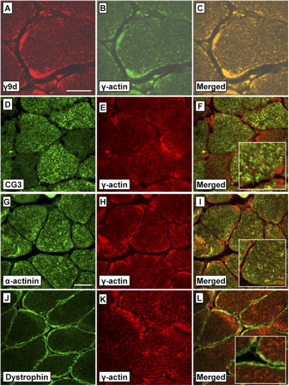
Tropomyosin (Tm) is a key component of the actin cytoskeleton and >40 isoforms have been described in mammals. In addition to the isoforms in the sarcomere, we now report the existence of two nonsarcomeric (NS) isoforms in skeletal muscle. These isoforms are excluded from the thin filament of the sarcomere and are localized to a novel Z-line adjacent structure. Immunostained cross sections indicate that one Tm defines a Z-line adjacent structure common to all myofibers, whereas the second Tm defines a spatially distinct structure unique to muscles that undergo chronic or repetitive contractions. When a Tm (Tm3) that is normally absent from muscle was expressed in mice it became associated with the Z-line adjacent structure. These mice display a muscular dystrophy and ragged-red fiber phenotype, suggestive of disruption of the membrane-associated cytoskeletal network. Our findings raise the possibility that mutations in these tropomyosin and these structures may underpin these types of myopathies.


Figures

Figure 1.
Figure 1.
Exon composition of tropomyosin (Tm) isoforms. The α-TM (A), β-TM (B), and γ-TM (C) gene organization is shown, with boxes representing exons, lines introns, and unshaded parts of the exons untranslated regions. Homologous exons of the three genes are shown in the same shading pattern. Constitutive exons that are present in all known isoforms are represented by solid black boxes. A vertical bar and the letter A represent alternate polyadenylation sites. Antibodies used in this study with the specific exons they detect are shown in italics on the gene maps. All possible isoforms from the three genes are not depicted. The 311 antibody detects all isoforms containing exon 1a which includes sarcomeric (αTmfast, βTm, and αTmslow) and NS Tms, and the α9d antibody detects 9d containing isoforms from the α-TM and β-TM genes. The γ-TM gene-specific antibodies CG3 and γ9d recognize isoforms containing exons 1b and 9d, respectively.
Figure 2.
Figure 2.
Expression of NS γ-TM products and nonmuscle actins in skeletal muscles. Shown are representative Western blots of protein (10 μg) from a range of mouse skeletal muscles and brain using antibodies that detect NS Tms from the γ-TM gene (A and B), γ-actins (C), and β-actin (D). The CG3 antibody (A) specifically recognizes the γ-TM 1b exon and detects all known NS products from this gene, whereas the γ9d antibody (B) recognizes a subset of these NS Tms, those that contain exon 9d (Fig. 1 C). A novel Tm isoform was detected at high levels in a subset of the muscles examined (EOM, soleus, and diaphragm), a 34-kD protein from γ-TM containing exon 1b (A, CG3 antibody). This novel isoform has not been detected in nonmuscle tissues. Both γ- and β-actin were detected in all muscles (C and D, respectively), but the levels were far greater for the former versus later isoforms. (Note that exposure to film was 45 s for γ-actin vs. 20 min for β-actin.) The γ-actin antibody also detects γ-smooth actin and so some of the signal with this antibody may be this isoform. (E) A representative Coomassie-stained gel used for the Westerns showing equal protein loading for the muscle extracts. Muscle abbreviations: ECU, extensor carpi ulnaris; EDL, extensor digitorum longus; EOM, extraocular muscles; FDP, flexor digitorum profundus; gastroc, gastrocnemius; quad, quadriceps; diaph, diaphragm.
Figure 3.
Figure 3.
Tm5NM-34kd is expressed in slow fibers, whereas the γ9d isoforms are expressed in all major fiber types. Consecutive transverse sections (7 μm) through the soleus muscle shows that fibers that were positive for the CG3 antibody (A and B, asterisks) were slow fibers (stained with the slow myosin heavy chain (MHC) antibody in B. Sections were stained with the primary antibodies followed by peroxidase-conjugated goat/anti–mouse secondary antibodies. In contrast to CG3, γ9d stained all fibers in soleus (C) and EDL (E) muscles. Note that between them the soleus and EDL contain all adult skeletal muscle fiber types (types I, IIA, IIX, and IIB). Negative (−ve) control sections (D and F) with the secondary antibody (donkey anti–sheep Alexa 488) confirms the specificity of the staining with γ9d. Bars, 40 μm.
Figure 4.
Figure 4.
The NS Tms from the γ-gene and a γ-actin are colocalized to a novel Z-line adjacent region of the sarcomere. Confocal immunofluorescent images of semi-thin (0.5–1.0 μm) longitudinal sections through adult soleus muscle show that Tms detected by CG3 and γ9d (A, C, D, F, G, and I, red signal) are localized to a restricted area either side of Z-line (B, C, H, and I, delineated by the green α-actinin staining), but not including the Z-line. The restricted localization of these NS Tms to either side of the Z-line is in marked contrast to the broad region of the actin thin filament of the sarcomere (E and F, phalloidin). In longitudinal sections there appears to be little difference in the Z-line adjacent localization of the Tms detected by CG3 and γ9d (J–L). Antibodies to a γ-actin also stained the Z-line adjacent region (M–O) and this staining was coincident with the staining for both CG3 (P–R) and γ9d (S–U). In some sections, γ-actin staining was observed at the Z-line (M and O, arrowheads; R, inset). Double staining of muscle sections was performed by applying both the primary/secondary antibody pairs sequentially. Bars, 2.5 μm.
Figure 5.
Figure 5.
NS Tms recognized by the CG3 and γ9d antibodies have distinct myofiber localization. Shown are confocal images of transverse sections (7 μm) through soleus muscles stained with γ9d (A) or CG3 (D) and costained with α-actinin (B and E). The enlarged merged images (C and F, insets) indicate that the Tms recognized by γ9d and CG3 are not colocalized with α-actinin (labels myofibrils) and therefore are located outside the myofibrils. Further transverse sections (G–I) show that CG3 and γ9d stain separate regions within the myofiber (particularly notable in the enlarged inset, I). Cross sections costained with the membrane protein dystrophin (J) and γ9d (K) also show strong staining of γ9d at the myofiber periphery beneath the membrane (L, arrow in the enlarged inset). Bars, 20 μm.
Figure 6.
Figure 6.
In transverse section, the γ9d Tms localize with a γ-actin, whereas the CG3 Tms do not. Shown are confocal images of transverse sections (7 μm) through soleus muscles stained with γ9d (A) or CG3 (D) and costained with a γ-actin antibody (B and E). The merged images show that the Tms recognized by γ9d colocalize with a γ-actin within the myofiber and at the fiber periphery (C), whereas the CG3 Tms are not localized with γ-actin (F). In addition, signal for a γ-actin did not coincide with α-actinin (I, enlarged inset) indicating that γ-actin microfilaments are not closely associated with the myofibrils. The most intense γ-actin staining was at the myofiber periphery (B, E, H, and K), but the staining was mostly distinct from the sarcolemmal staining of dystrophin (L, inset). Bars, 20 μm.
Figure 7.
Figure 7.
Expression of high levels of Tm3 in muscle has little effect on expression of sarcomeric and NS Tms. Western blots of 6-mo-old Tm3 skeletal muscle showing the influence of high levels of Tm3 expression on (A) NS or (B) sarcomeric Tm isoforms. Results for both wild-type (WT) and high-expressing transgenic lines (3/70 and 3/66) are shown. NS Tms containing exon 9d from the α- and β-TM genes are recognized by α9d. Products containing exon 1b and exon 9d from the γ-TM gene are recognized by CG3 and γ9d, respectively. A Tm3 band is detected by the γ9d antibody in Tm3 mice (3/70 and 3/66) due to cross-reactivity of the antibody with exon 9d products from the α-TM gene. The 311 antibody recognizes Tms containing exon 1a (Fig. 1), which includes Tm3 and the three sarcomeric Tms (αTmfast, αTmslow, and βTm). Under the electrophoretic conditions used the αTmfast and αTmslow isoforms are seen as a single band. See Fig. 2 legend for muscle abbreviations.
Figure 8.
Figure 8.
Ectopically expressed Tm3 accumulates adjacent to the Z-line and at the M-line. Shown are immunofluorescent confocal images of longitudinal and transverse sections of soleus muscle from adult Tm3 transgenic (line 3/66) and wild-type (WT) mice. WT soleus muscle contains two main Tm isoforms (Tm1 and Tm6; Fig. 7 A) recognized by the α9d antibody. Longitudinal and transverse sections (7 μm) of WT muscle indicate that these isoforms are present in structures outside of the myofiber and at the myofiber periphery (D and J, respectively). Tm3 (also detected by the α9d antibody; Fig. 1 A), is absent from WT muscle (Fig. 7 A), but in the Tm3 mice the ectopic protein is located at the Z-line adjacent region (A–C, 0.5–1.0 μm longitudinal sections; Z-line marked by arrows) and at the M-line (A and C, arrowheads). Tm3 localized mainly to the sarcomeric compartment (G and K) and was colocalized with the γ-actin antibody in longitudinal and cross sections (I and N, respectively). Bars: (A–C) 2.5 μm; (D–I) 40 μm; and (J–N) 20 μm.
Figure 9.
Figure 9.
Inappropriate expression of an NS Tm (Tm3) results in muscular dystrophy phenotype and ragged-red fibers. The Tm3 mice have a number of features characteristic of muscular dystrophy in quadriceps (A–C) and soleus muscles (D–F). Features characteristic of muscular dystrophy shown in H&E stained transverse sections (A–F) include: areas of regenerating fibers with centralized nuclei (B, C, and F, black arrows), myofiber size variability (B, C, and F), and regions of fiber degeneration (F, white arrow), macrophage infiltration (B and F, black double arrows) and fibrosis (F, white arrowhead). Soleus muscle from Tm3 mice also has the characteristic accumulation of sarcolemmal mitochondria that is associated with ragged red fibers. Mitochondrial accumulations (black arrowheads) were observed in Gomori-Trichrome (G) and H&E stained (E) sections in of soleus of line 3/66 mice. The large increase in number and size of mitochondria beneath the sarcolemma is clearly evident in electron micrographs from Tm3 mice (J) compared with WT mice (I). Asterisks in I and J mark the subsarcolemmal mitochondria. Bars, A–H, 40 μm. When suspended by the tail the Tm3 mice (line 3/66; L), unlike the WT mice (K), are unable to extend their hindlimbs away from the body. This phenomenon has been observed in a mouse model of muscular dystrophy (Bittner et al., 1999).
Figure 10.
Figure 10.
Schematic representation of NS Tm localization in skeletal muscle. Depicted are myofibers in longitudinal (A) and transverse section (B) showing the location of the NS Tms and γ-actin in relation to the major structures in skeletal muscle. (A) In the longitudinal representation, two main membrane/cytoskeletal junctions are depicted: (1) integrin-based focal adhesions and (2) the dystrophin glycoprotein complex. Hypothesized tropomyosin-associated γ-actin networks that link the sarcomeric arrays to the costamere are shown as black lines. Other filaments linking the M-line and Z-line to the membrane are not depicted. The results of this study suggest that the NS tropomyosins recognized by the γ9d and CG3 antibodies (Tm5NM1 and Tm5NM-34kd, respectively) are located adjacent to the Z-line in the sarcomeric space. Data suggest that Tm5NM1 associates with a γ-actin whereas the actin (broken black line) interacting with Tm5NM-34kd was not defined. Note that although α-actinin (pink) is only shown cross-linking γ-actin filaments, its main location is in the Z-line linking sarcomeric actin thin filaments from adjacent sarcomeres. (B) In the transverse representation, Tm5NM1 (blue) and Tm5NM-34kd (green) are shown in the intermyofibrillar space, the former in association with a γ-actin and the later with an unknown actin filament system. Also shown is Tm5NM1/γ-actin filaments at the subsarcolemmal region, a space also occupied by mitochondria. The data suggests that the Tm5NM1 filaments are in all adult fibers, whereas the Tm5NM-34kd structure is restricted to specialized subset of fibers. The ectopic Tm3 localizes to the Z-line–associated γ-actin cytoskeleton and results in muscular dystrophy and ragged-red fibers.

Similar articles

Cited by

References

    1. Arai, A., J.A. Spencer, and E.N. Olson. 2002. STARS, a striated muscle activator of Rho signaling and serum response factor-dependent transcription. J. Biol. Chem. 277:24453–24459. - PubMed
    1. Bang, M.L., R.E. Mudry, A.S. McElhinny, K. Trombitas, A.J. Geach, R. Yamasaki, H. Sorimachi, H. Granzier, C.C. Gregorio, and S. Labeit. 2001. Myopalladin, a novel 145-kilodalton sarcomeric protein with multiple roles in Z-disc and I-band protein assemblies. J. Cell Biol. 153:413–427. - PMC - PubMed
    1. Banker, B.Q., and A.G. Engel. 1994. Basic Reactions of Muscle. Myology. A.G. Engel and Franzini-Armstrong C., editors. McGraw-Hill, Inc., New York. 832–888.
    1. Bittner, R.E., L.V. Anderson, E. Burkhardt, R. Bashir, E. Vafiadaki, S. Ivanova, T. Raffelsberger, I. Maerk, H. Hoger, M. Jung, et al. 1999. Dysferlin deletion in SJL mice (SJL-Dysf) defines a natural model for limb girdle muscular dystrophy 2B. Nat. Genet. 23:141–142. - PubMed
    1. Borrione, A.C., A.M. Zanellato, L. Saggin, M. Mazzoli, G. Azzarello, and S. Sartore. 1988. Neonatal myosin heavy chains are not expressed in Ni-induced rat rhabdomyosarcoma. Differentiation. 38:49–59. - PubMed

Publication types

MeSH terms