Comparative Study
. 2004 Nov;87(5):3460-9.
doi: 10.1529/biophysj.104.044024. Epub 2004 Aug 31.

Analysis of side-chain rotamers in transmembrane proteins

Affiliations
Comparative Study

Analysis of side-chain rotamers in transmembrane proteins

Aaron K Chamberlain et al. Biophys J. 2004 Nov.

Abstract

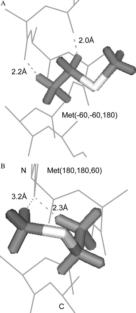
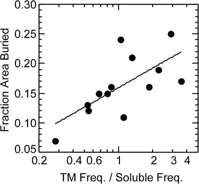
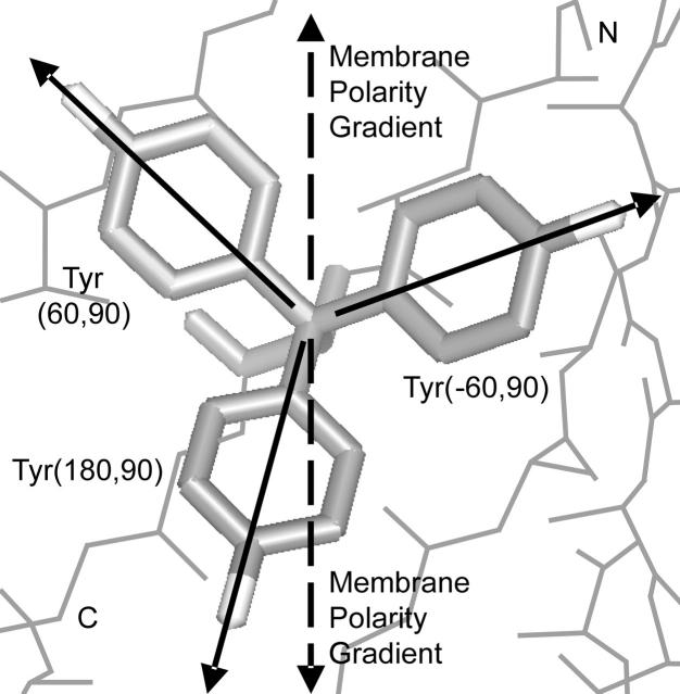
We measured the frequency of side-chain rotamers in 14 alpha-helical and 16 beta-barrel membrane protein structures and found that the membrane environment considerably perturbs the rotamer frequencies compared to soluble proteins. Although there are limited experimental data, we found statistically significant changes in rotamer preferences depending on the residue environment. Rotamer distributions were influenced by whether the residues were lipid or protein facing, and whether the residues were found near the N- or C-terminus. Hydrogen-bonding interactions with the helical backbone perturbs the rotamer populations of Ser and His. Trp and Tyr favor side-chain conformations that allow their side chains to extend their polar atoms out of the membrane core, thereby aligning the side-chain polarity gradient with the polarity gradient of the membrane. Our results demonstrate how the membrane environment influences protein structures, providing information that will be useful in the structure prediction and design of transmembrane proteins.


Figures

FIGURE 1
FIGURE 1
The three favored χ1 angles in proteins. The −60°, +60°, and 180° angles are often referred to as gauche minus (g−), gauche plus (g+), and trans (t), respectively. We refer to a rotamer by the amino acid name followed by the χ-angles in the center of the rotamer bins, e.g., Leu(−60,180).
FIGURE 2
FIGURE 2
Two Trp rotamers allowing extension of ring N atom toward the N- and C-terminal sides of the membrane. Trp(−60,120) extends the N atom 2.6 Å toward the N-terminal side of the membrane whereas Trp(180,0) extends 2.2 Å toward the C-terminal side. The Cβ atom is considered the reference point.
FIGURE 3
FIGURE 3
Histidine's most frequent rotamer in TM helices, His(−60,60). The side-chain Nδ atom hydrogen bonds to the i − 4 backbone carbonyl O atom. The Nδ–O distance is 2.2 Å. This rotamer also extends the ring N atoms an average of 2.0 Å toward the N-terminal side of the membrane.
FIGURE 4
FIGURE 4
The Phe(−60,90) rotamer making two potential C-H⋯O hydrogen bonds to backbone O atoms in TM helices. The dashed lines indicate the distances from the Hβ and Hδ atoms to the i − 3 and i − 4 carbonyl O atoms, respectively.
FIGURE 5
FIGURE 5
Weak hydrogen-bonding interactions in Met rotamers in TM helices. (A) The six Met(−60,−60 or 180,X) rotamers can make two weak hydrogen bonds to carbonyl O atoms. Met(−60,−60,180) is shown with the two C-H⋯O bonds indicated by dotted lines. (B) The second most-frequent Met rotamer in TM helices, Met(180,180,60), has two potential C-H⋯O bonds. One bond, the Hβ-O bond, is the same as in panel A, but the other is a novel bond using the methyl Hɛ atom.
FIGURE 6
FIGURE 6
The surface area buried and change in rotamer frequency of Met rotamers in TM helices. The fraction of surface area buried is shown as a function of the increase in rotamer frequency in TM helices compared to soluble helices. Numbers larger than one on the abscissa indicate that the TM rotamer frequency is higher than the soluble rotamer frequency. Points are plotted for the 13 rotamers with a frequency >1% in soluble proteins.
FIGURE 7
FIGURE 7
The three most-frequent Tyr rotamers in TM barrels. Each rotamer is marked by a solid vector from low to high polarity. The polarity gradients of the membrane extend out of the membrane toward either the N- or C-terminal side (dashed vectors). Tyr(180,90) aligns it polarity gradient better than the other Tyr rotamers with one of the membrane polarity gradients. This rotamer extends toward the C-terminus helping Tyr to be more populated on this side of the membrane.

References

    1. Andersson, H., and G. von Heijne. 1994. Membrane protein topology: effects of delta mu H+ on the translocation of charged residues explain the “positive inside” rule. EMBO J. 13:2267–2272. - PMC - PubMed
    1. Arkin, I. T., and A. T. Brunger. 1998. Statistical analysis of predicted transmembrane alpha-helices. Biochim. Biophys. Acta. 1429:113–128. - PubMed
    1. Bowie, J. U., R. Luthy, and D. Eisenberg. 1991. A method to identify protein sequences that fold into a known three-dimensional structure. Science. 253:164–170. - PubMed
    1. Burley, S. K., and G. A. Petsko. 1988. Weakly polar interactions in proteins. Adv. Protein Chem. 39:125–189. - PubMed
    1. Chakrabarti, P., and S. Chakrabarti. 1998. C–H…O hydrogen bond involving proline residues in alpha-helices. J. Mol. Biol. 284:867–873. - PubMed

Publication types

LinkOut - more resources