. 2004 Nov 12;279(46):48177-88.
doi: 10.1074/jbc.M407836200. Epub 2004 Aug 31.

Mutational analysis of arginine 276 in the leucine-loop of human uracil-DNA glycosylase

Affiliations

Mutational analysis of arginine 276 in the leucine-loop of human uracil-DNA glycosylase

Cheng-Yao Chen et al. J Biol Chem. .

Abstract

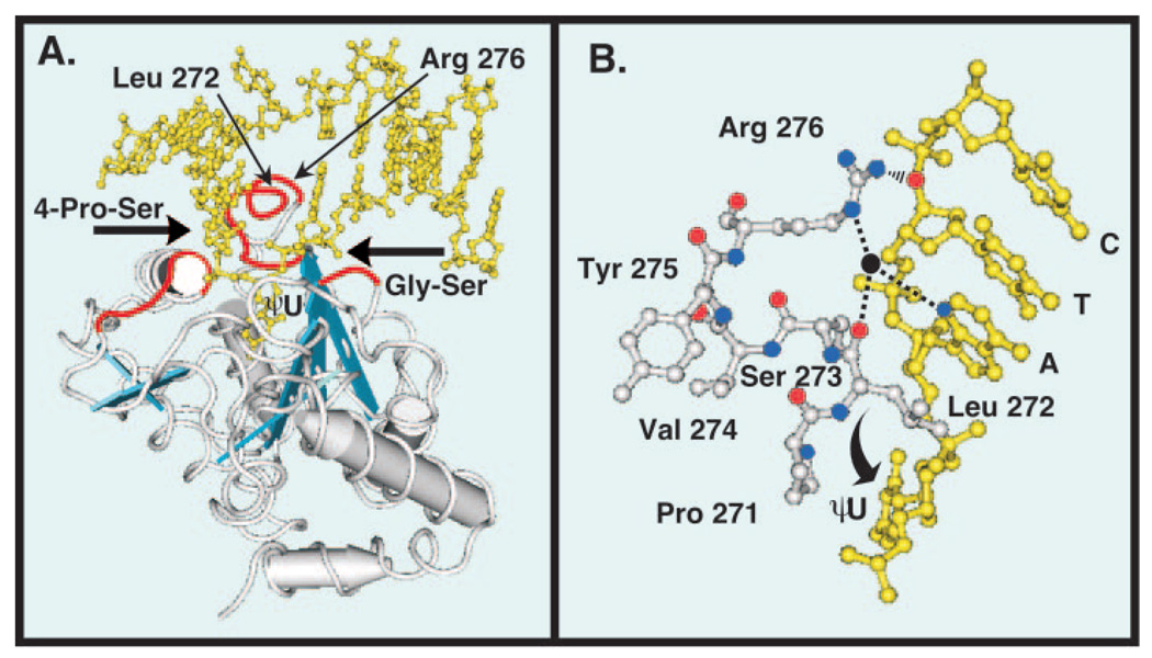
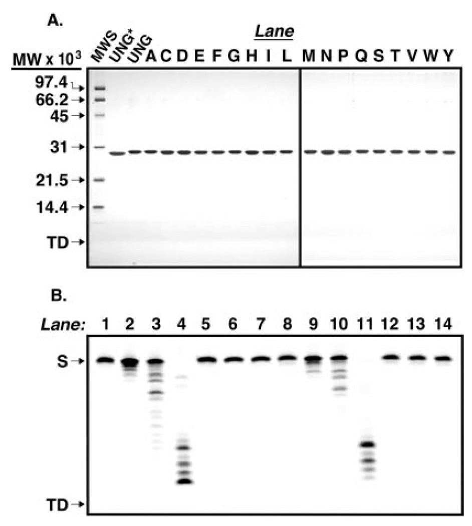
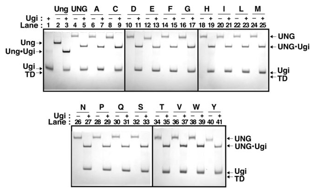
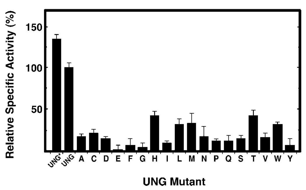
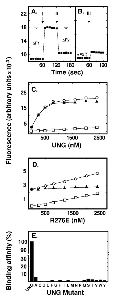
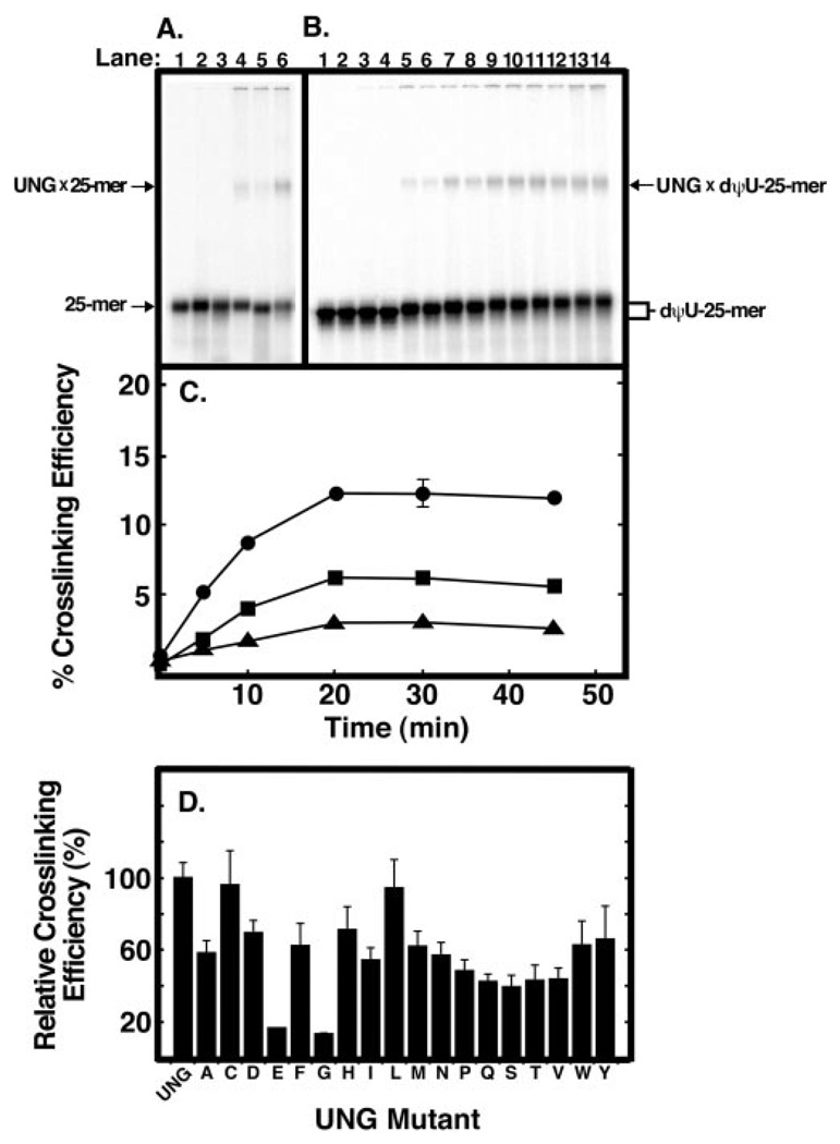
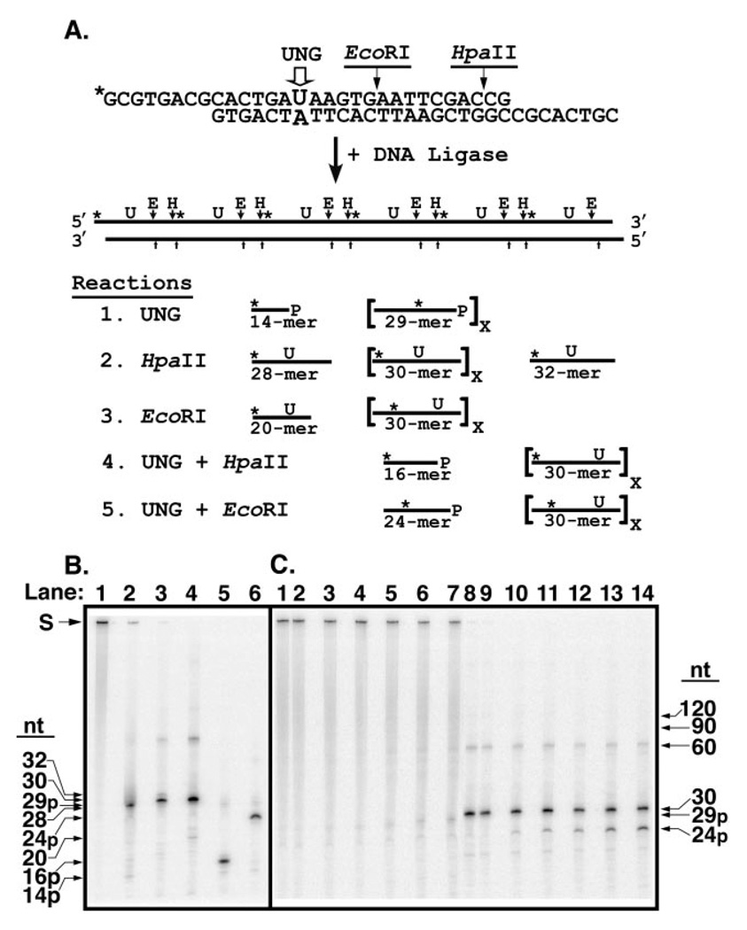
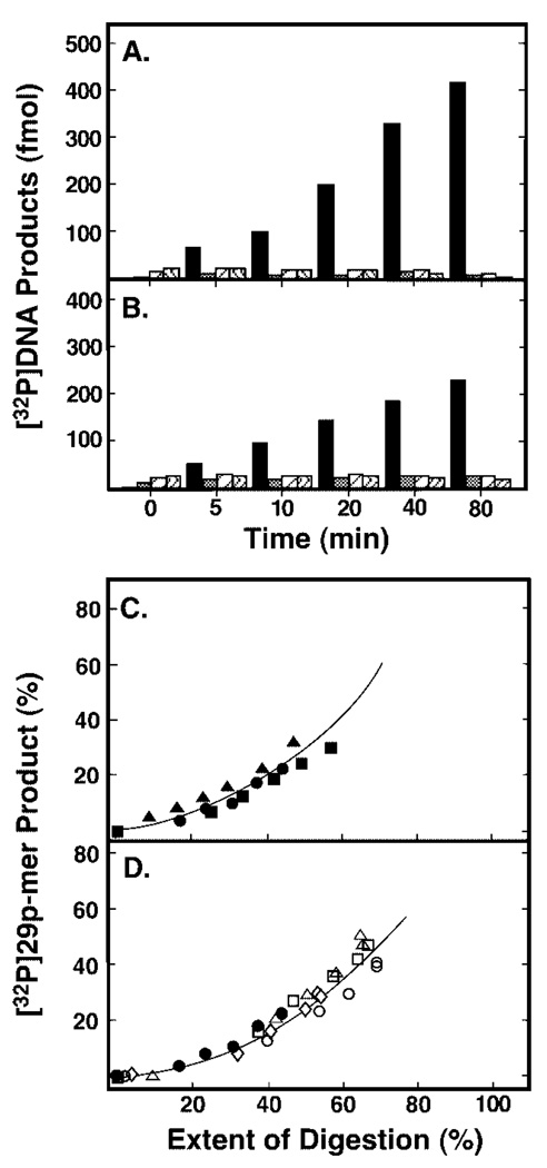
Uracil residues are eliminated from cellular DNA by uracil-DNA glycosylase, which cleaves the N-glycosylic bond between the uracil base and deoxyribose to initiate the uracil-DNA base excision repair pathway. Co-crystal structures of the core catalytic domain of human uracil-DNA glycosylase in complex with uracil-containing DNA suggested that arginine 276 in the highly conserved leucine intercalation loop may be important to enzyme interactions with DNA. To investigate further the role of Arg(276) in enzyme-DNA interactions, PCR-based codon-specific random mutagenesis, and site-specific mutagenesis were performed to construct a library of 18 amino acid changes at Arg(276). All of the R276X mutant proteins formed a stable complex with the uracil-DNA glycosylase inhibitor protein in vitro, indicating that the active site structure of the mutant enzymes was not perturbed. The catalytic activity of the R276X preparations was reduced; the least active mutant, R276E, exhibited 0.6% of wildtype activity, whereas the most active mutant, R276H, exhibited 43%. Equilibrium binding studies utilizing a 2-aminopurine deoxypseudouridine DNA substrate showed that all R276X mutants displayed greatly reduced base flipping/DNA binding. However, the efficiency of UV-catalyzed cross-linking of the R276X mutants to single-stranded DNA was much less compromised. Using a concatemeric [(32)P]U.A DNA polynucleotide substrate to assess enzyme processivity, human uracil-DNA glycosylase was shown to use a processive search mechanism to locate successive uracil residues, and Arg(276) mutations did not alter this attribute.


Figures

Fig. 1
Fig. 1. Tertiary structure of human uracil-DNA glycosylase bound to DNA
Co-crystal structure of the core catalytic domain UNG* bound to DNA containing the uncleaved uracil analogue dψU (5). The DNA is shown in yellow and the view is looking into the major groove. A, three distinct amino acid sequences (red tubes) of the UNG* polypeptide backbone (silver tubes) critical to the proposed pinch-pull-push catalytic mechanism (7) are shown. The 4 Pro-Ser loop (165PPPPS169) and the Gly-Ser loop (246GS247) compress (“pinch”) the deoxyribose phosphate backbone from the 5′ and 3′ directions, respectively (6). The Leu272 loop (268HPSPLSVYR276), which contains Arg276, penetrates the DNA base stack (“push”) and occupies the helical space of the flipped-out ψU residue (6). Conserved amino acid residues (Gln144, Asn204, and His268) in the UNG* binding pocket capture (“pull”) and stabilize the expelled extrahelical dψU. α-Helices are depicted as silver cylinders and β-sheets are illustrated as blue strands. B, ball-and-stick diagram of the UNG Leu272 loop (271PLSVYR276) shown in silver and a portion of the oligonucleotide sequence 3′-CTA dψU-5′ shown in yellow. The ηN of the Arg276 guanidinium side chain (nitrogen atoms, blue balls) is shown as interacting (black rippled lines) with the 5′-phosphate of the cytosine residue (oxygen atom, red ball), as stated for cleaved U·G DNA by Slupphaug et al. (17). The εN participates in water-bridged (water, black ball) hydrogen bonding (dashed lines) with the N3 of adenine (blue ball) and the carbonyl group (red ball) of Leu272 as shown in Parikh et al. (6). Structures were drawn with the Cn3D 4.0 software program using Protein Data Bank code 1EMH (MMDB 13471) deposited by Parikh et al. (5) in the Molecular Modeling Data base of the National Center for Biotechnology Information.
Fig. 2
Fig. 2. Purity of the enzymes used in this study
A, UNG* (fraction VIII), UNG, and various Arg276 mutant proteins (fraction IV) were purified as described under “Experimental Procedures.” Samples (4 µg) of UNG*, UNG, and each Arg276 mutant protein were subjected to 12.5% SDS-polyacrylamide gel electrophoresis, and protein bands were visualized after staining with Coomassie Brilliant Blue G-250. The mobility of molecular weight standards (SDS-PAGE, low-range standards, Bio-Rad) as well as the tracking dye (TD) are indicated by arrows from top to bottom, respectively (lane MWS). Lanes containing R276X mutant proteins are represented by conventional single letter amino acid abbreviations. B, UNG (fraction IV) was assayed for nuclease activity using 5′-end carboxyfluorescein-labeled single- and double-stranded oligonucleotide substrates, 5′-FAM-T-25-mer and 5′- FAM-T·A-25-mer, respectively, as described under “Experimental Procedures.” Mock reaction mixtures contained 5′-FAM-T-25-mer (12.5 ng) or 5′-FAM-T·A-25-mer (25 ng) in reaction buffer (lanes 1 and 8, respectively), control reactions contained 0.02, 0.2, or 2 units of E. coli exonuclease III and either 5′-FAM-T-25-mer or 5′-FAM-T·A-25-mer (lanes 2–4 and 9–11, respectively), and reactions containing UNG (8, 40, and 200 ng) and 5′-FAM-T-25-mer or 5′-FAM-T·A-25-mer (lanes 5–7, and 12–14, respectively) were incubated at 37 °C for 30 min. The reaction products were resolved by 15% polyacrylamide, 8.3 m urea, gel electrophoresis, and the gel was analyzed with a FMBioII fluorescence imaging system. Arrows indicate the locations of oligonucleotide substrate (S) and tracking dye (TD).
Fig. 3
Fig. 3. Ability of UNG and R276X mutant proteins to bind Ugi
Reaction mixtures (15 µl) containing 40 pmol of E. coli Ung (lanes 2 and 3), UNG (lanes 4 and 5), or R276X mutant protein (lanes 6–41), with or without (+ or −) Ugi (100 pmol) were incubated as described under “Experimental Procedures.” A control reaction containing Ugi (100 pmol) alone was similarly processed (lane 1). Samples were analyzed at 4 °C by non-denaturing 10% polyacrylamide gel electrophoresis, proteins were visualized with Coomassie Brilliant Blue G-250 stain, and the gel was imaged as described under “Experimental Procedures.” Arrows indicate the location of Ung, UNG, Ugi, Ung·Ugi, UNG·Ugi, and the tracking dye front (TD). Arg276 mutant protein lane assignments are indicated by single letter amino acid abbreviations.
Fig. 4
Fig. 4. Specific uracil-DNA glycosylase activity of R276X mutant proteins
Uracil-DNA glycosylase activity was measured under standard reaction conditions using 0.04–0.13 units of UNG*, UNG, or the indicated R276X mutant protein, as described under “Experimental Procedures.” Protein concentrations were determined using the Bradford method (36) and the Protein Assay reagent (Bio-Rad). The specific activity (units/mg) of UNG* and the R276X mutant enzymes was normalized to that of UNG (3.76 × 10⁵ units/mg), which was defined as 100%. Arg276 mutant proteins are denoted by single letter amino acid abbreviations. Error bars represent the standard deviation of four experimental determinations.
Fig. 5
Fig. 5. Effect of Arg276 mutations on DNA binding and base flipping as measured by 2-aminopurine fluorescence
Reaction mixtures (250 µl) contained 50 nm double-stranded dψU·2AP-25-mer and various amounts (0–1500 nm) of UNG or Arg276 mutant proteins. Fluorescence intensity measurements were performed as described under “Experimental Procedures.” A, the baseline fluorescent intensity of the 2-aminopurine-containing (dψU·2AP-25-mer) DNA substrate was measured at 1-s intervals for 1 min. UNG (500 nm) was then added and additional fluorescent intensity measurements were continued for 1 min. Arrow I marks the time of UNG addition; ΔF1 is the average fluorescence intensity enhancement caused by UNG addition. Lastly, Ugi (5 µm) was added to the reaction and the fluorescence intensity was monitored for another minute. Arrow II marks the time of Ugi addition and ΔF2 is the average 2AP fluorescent intensity quench that resulted from Ugi addition. The rectangular plot symbols represent the average intensity of 10 consecutive 1-s measurements. B, the fluorescent intensity of the dψU·2AP-25-mer was monitored for 1 min as in A prior to the addition of Ugi (5 µm). Arrow III marks the time of Ugi addition; ΔF3 corresponds to the average increase in 2AP fluorescent intensity caused by Ugi addition. C, reaction mixtures containing dψU·2AP-25-mer (50 nm) and 0, 100, 250, 500, 1000, or 1500 nm UNG were prepared and the fluorescent intensity was determined at 1-s intervals for 1 min. Open circles represent total observed fluorescence, open squares represent the fluorescence of the protein solution alone, and filled triangles represent the net fluorescence (open circles minus open squares). D, samples were prepared as described in C, except that the R276E mutant enzyme replaced UNG. The linear dependence of fluorescent intensity as a function of the enzyme concentration was calculated and termed the initial “slope” of the binding curve. Plot symbols are as described in C. E, sample preparation, fluorescent intensity measurements, and slope calculations were carried out as described in D for each of the Arg276 mutant enzyme preparations and compared with the initial slope of UNG, which was set to 100%. Error bars indicating the standard deviation of the 60 1-s fluorescent intensity measurements in C–E are obscured by the plot symbols.
Fig. 6
Fig. 6. Ability of UNG and Arg276 mutant proteins to form UV-catalyzed cross-links to [32P]25-mer DNA
A, samples (12 µl) containing 20 pmol of 5′-end 32P-labeled oligonucleotide T-25-mer, U-25-mer, or dψU-25-mer without UNG (lanes 1–3, respectively) or with 40 pmol of UNG (lanes 4–6, respectively) were UV-irradiated for 30 min as described under “Experimental Procedures.” Following irradiation, samples were subjected to non-denaturing polyacrylamide gel electrophoresis; the gels were dried and analyzed using a PhosphorImager. The positions of the UNG × [32P]DNA-25-mer cross-linked complex bands and free [32P]DNA-25-mer bands are indicated by arrows. B, reaction mixtures (12 µl) were prepared in duplicate that contained 20 pmol of [32P]dψU-25-mer and 40 pmol of UNG. Following UV irradiation for 0, 5, 10, 20, 30, and 45 min (lanes 3–14, respectively), reactions were analyzed as in A. Control reactions (lanes 1 and 2), containing 20 pmol of [32P]dψU-25-mer, were not irradiated. C, reaction mixtures (12 µl) were prepared in duplicate that contained 20 pmol of [32P]dψU-25-mer (closed circles), [32P]T-25-mer (closed squares), or [32P]U-25-mer (closed triangles), and 40 pmol of UNG. Following UV irradiation for 0, 5, 10, 20, 30, and 45 min, reactions were analyzed as in A, and the PhosphorImager data were quantified using the ImageQuant program. The cross-linking efficiency (%) was calculated by dividing the intensity of the UNG × [32P]25-mer band by the sum of the [32P]25-mer and UNG × [32P]25-mer bands and multiplying by 100. D, reaction mixtures (12 µl) were prepared that contained 20 pmol of [32P]dψU-25-mer and 40 pmol of each Arg276 mutant enzyme. The reactions were UV-irradiated for 10 min and analyzed as described in C. The cross-linking efficiency of each mutant preparation, indicated by the corresponding single letter amino acid abbreviation, is compared with that of UNG. Error bars represent the standard deviation of three experiments.
Fig. 7
Fig. 7. Analysis of reaction products generated by UNG from a concatenated uracil-containing [32P]DNA substrate
A, the concatenated uracil-containing [32P]DNA substrate was prepared by ligation of double-stranded 30-mer units as described under “Experimental Procedures.” Each 30-mer unit contained a uracil residue at position 15 in the upper strand opposite adenine in the lower, complementary, DNA strand; only the uracil-containing strand was 5′-32P-end labeled (marked with an asterisk, *). An EcoRI restriction endonuclease recognition site was situated 3′ to nucleotide 20 of the uracil-containing DNA strand. The 30-mer unit was designed with self-complementary overhanging 5′-ends of eight nucleotides that formed a HpaII endonuclease recognition site upon ligation. Ligation of the 30-mer units created a concatenated uracil-containing [32P]DNA substrate in which each uracil residue as well as the EcoRI and HpaII recognition sites were separated by 30 nucleotides. Reaction 1: sequential uracil excision by UNG followed by alkaline hydrolysis of the resultant AP-site produces 29-mers containing an internal 32P label and a 3′-phosphate (29p-mer). Excision of the 5′-terminal uracil produces a 5′-32P-end labeled 14-mer with a 3′-phosphate (14p-mer). Reaction 2: HpaII digestion of the concatenated [32P]DNA substrate and subsequent alkaline hydrolysis generates 30-mers with an internal 32P label. Cleavage by HpaII at the 5′- and 3′-ends yields a 28-mer containing a 5′-32P-end label and a 32-mer containing an internal 32P label, respectively. Reaction 3: EcoRI digestion produces 30-mers except at the 5′ terminus where a [32P]DNA-20-mer is generated. Reaction 4: concatenated [32P]DNA digested with UNG, then exhaustively digested with HpaII followed by alkaline hydrolysis, yields 16-mers containing a 3′-phosphate (16p-mer) where the uracil has been excised, but [32P]30-mers from intact uracil-containing 30-mer units, as shown in Reaction 2. Only 32P-labeled reaction products are shown. Reaction 5: concatenated [32P]DNA digested with UNG, then exhaustively digested with EcoRI followed by alkaline hydrolysis, yields [32P]24-mers containing a 3′-phosphate (24p-mer) where the uracil has been excised, but [32P]30-mers from uracil-containing 30-mer units, as shown in Reaction 3. Only 32P-labeled reaction products are shown. B, characterization of the concatenated uracil-containing [32P]DNA substrate. Reaction mixtures (10 µl) containing 10 pmol of concatenated [32P]DNA and no addition (lane 1), 70 units of UNG (lane 2), 10 units of EcoRI (lane 3), 10 units of HpaII (lane 4), 70 units of UNG and 10 units of HpaII (lane 5), or 70 units of UNG and 10 units of EcoRI (lane 6), were incubated at 37 °C, subjected to alkaline hydrolysis, and analyzed by 12% denaturing polyacrylamide gel electrophoresis as described under “Experimental Procedures.” The mobility of the unreacted concatenated [32P]DNA substrate (S) and various oligonucleotide reaction products (32-, 30-, 29p-, 28-, 24p-, 20-, 16p-, and 14p-mer) is indicated by arrows. C, time course of UNG digestion. A reaction mixture (100 µl) containing 0.06 units of UNG and 80 pmol of concatenated uracil-containing [32P]DNA was incubated at 37 °C. Aliquots (10 µl) were removed at 0, 5, 10, 20, 40, and 80 min and terminated by the addition of Ugi as described under “Experimental Procedures.” A control reaction contained 10 pmol of the [32P]DNA substrate only. Following reaction termination, each time point was divided into two aliquots. One, designated the UNG reaction, was subjected to alkaline hydrolysis. The other, designated as the EcoRI reaction, underwent EcoRI digestion and subsequent alkaline hydrolysis. Reactions were analyzed by denaturing 12% polyacrylamide, 8.3 m urea gel electrophoresis as described under “Experimental Procedures.” UNG reactions corresponding to the control reaction, and 0-, 5-, 10-, 20-, 40-, and 80-min time points were loaded in lanes 1–7, respectively, whereas the corresponding EcoRI reactions were loaded in lanes 8–14. Arrows indicate the positions of the various oligonucleotide reaction products. The extent of uracil excision after each incubation time was determined by dividing the amount of [32P]24p-mer oligonucleotide released by EcoRI digestion by the sum of [32P]24p-mer + [32P]30-mer, and multiplying the quotient by 100; this percentage was called the Extent of Digestion.
Fig. 8
Fig. 8. Analysis of reaction products generated by Ung, UNG*, UNG, or its Arg276 mutant proteins from concatenated uracil-containing [32P]DNA
Two processivity reaction mixtures (100 µl) containing either 0.04 units of E. coli Ung (A) or 0.06 units of human UNG* (B), and 80 pmol of concatenated uracil-containing [32P]DNA substrate were prepared as described under “Experimental Procedures.” Samples (10 µl) were withdrawn after 0, 5, 10, 20, 40, and 80 min of incubation at 37 °C, and divided into two aliquots as described in the legend to Fig. 7. From the reactions subjected to alkaline hydrolysis only, the molar quantities of the four detectable [32P]oligonucleotide UNG digestion products were determined and are presented graphically as: 29p-mer (black bar), 59p-mer (cross-hatched bar), 89p-mer (striped bar), and 119p-mer (wavy bar). C, the amount of [32P]29p-mer product generated at each incubation time (A and B, black bars) was determined relative to the total [32P]DNA detected, and the extent of digestion at various incubation times was determined after EcoRI treatment. A time course reaction identical to that described for Ung and UNG* but containing 0.06 unit of UNG was also conducted and analyzed as described above. The processivity of Ung (0.04 unit, filled circles), UNG* (0.06 unit, filled squares), or UNG (0.06 unit, filled triangles) was analyzed by graphing the amount (%) of processive [32P]29p-mer product detected as a function of the extent of digestion (%). D, processivity analysis of uracil-DNA glycosylase reactions containing R276D (0.3 unit, open diamonds), R276L (0.3 unit, open circles), R276T (1.0 unit, open triangles), and R276W (1.0 unit, open squares) is presented and compared with UNG (0.06 unit, filled circles).

Similar articles

Cited by

References

    1. Lindahl T, Ljungquist S, Siegert W, Nyberg B, Sperens B. J. Biol. Chem. 1977;252:3286–3294. - PubMed
    1. Aravind L, Koonin EV. Genome Biol. 2000;1:0007.0001–0007.0008. - PMC - PubMed
    1. Pearl LH. Mutat. Res. 2000;460:165–191. - PubMed
    1. Mosbaugh DW, Bennett SE. Prog. Nucleic Acids Res. Mol. Biol. 1994;48:315–370. - PubMed
    1. Parikh SS, Walcher G, Jones GD, Slupphaug G, Krokan HE, Blackburn GM, Tainer JA. Proc. Natl. Acad. Sci. U. S. A. 2000;97:5083–5088. - PMC - PubMed

Publication types

LinkOut - more resources