. 2004 Sep;24(18):7976-86.
doi: 10.1128/MCB.24.18.7976-7986.2004.

The small nucle(ol)ar RNA cap trimethyltransferase is required for ribosome synthesis and intact nucleolar morphology

Affiliations

The small nucle(ol)ar RNA cap trimethyltransferase is required for ribosome synthesis and intact nucleolar morphology

Geoffroy Colau et al. Mol Cell Biol. 2004 Sep.

Abstract

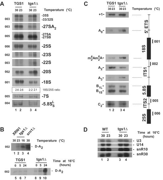
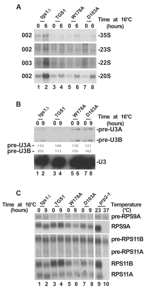
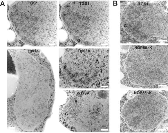
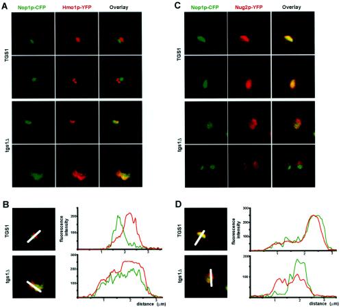
Nucleolar morphogenesis is a poorly defined process. Here we report that the Saccharomyces cerevisiae nucleolar trimethyl guanosine synthase I (Tgs1p), which specifically selects the m(7)G cap structure of snRNAs and snoRNAs for m(2,2,7)G conversion, is required not only for efficient pre-mRNA splicing but also for pre-rRNA processing and small ribosomal subunit synthesis. Mutational analysis indicates that the requirement for Tgs1p in pre-mRNA splicing, but not its involvement in ribosome synthesis, is dependent upon its function in cap trimethylation. In addition, we report that cells lacking Tgs1p showed a striking and unexpected loss of nucleolar structural organization. Tgs1p is not a core component of the snoRNP proteins; however, in vitro, the protein interacts with the KKD/E domain present at the carboxyl-terminal ends of several snoRNP proteins. Strains expressing versions of the snoRNPs lacking the KKD/E domain were also defective for nucleolar morphology and showed a loss of nucleolar compaction. We propose that the transient and functional interactions of Tgs1p with the abundant snoRNPs, through presumed interactions with the KKD/E domain of the snoRNP proteins, contribute substantially to the coalescence of nucleolar components. This conclusion is compatible with a model of self-organization for nucleolar assembly.


Figures

FIG. 1.
FIG. 1.
Tgs1p is required for small ribosomal subunit synthesis. (A) Yeast pre-rRNA processing pathway. Cleavage sites (A to E) and oligonucleotide probes (001 to 006) used in the Northern blot and primer extension experiments are indicated. See text and reference for a detailed description of the pathway. The box C+D snoRNAs U3 and U14 and the box H+ACA snoRNAs snR10 and snR30 are required for pre-rRNA processing at sites A0 to A2. U3, snR10, and snR30 are cap trimethylated by Tgs1p. ETS, external transcribed spacer. (B) In vivo labeling analysis of pre-rRNA processing in strains with TGS1 deleted (tgs1Δ) and a wild-type isogenic control (TGS1) grown at the semipermissive temperature of 23°C. Samples were collected after 1, 2, 5, and 10 min of chase.
FIG. 2.
FIG. 2.
Tgs1p is required for pre-rRNA processing. (A to C) Northern blot (A and B) and primer extension (C) analyses of pre-rRNA processing defects in tgs1Δ and isogenic wild-type backgrounds. Temperatures of growth (16, 23, and 30°C) and the oligonucleotides used (001 to 006 [Fig. 1A]) are indicated. The ratio of 18S to 25S was quantitated with a PhosphorImager (Typhoon; Amersham Biosciences) and estimated to be ∼2.6 for the wild type and ∼2.1 to ∼2.2 for the mutant. tgs1Δ cells grown under nonpermissive conditions accumulated the D-A2 spacer fragment that is readily detected in xrn1Δ strains (27) (B). The use of the two alternative pathways of 5.8S-25S synthesis (30) was not affected in tgs1Δ strains, as indicated by the steady-state levels of the two forms of 5.8S rRNA (A) and the conserved ratio of cleavage at site B1S and B1L (C). In wild-type cells, the 18S rRNA is dimethylated at its 3′ end on two adjacent adenosines by Dim1p (9). cDNA strands that extended across this position from oligonucleotide 002 revealed that pre-rRNAs accumulated in tgs1Δ cells are dimethylated (C). (D) Steady-state levels of the box C+D snoRNAs U3 and U14 and the box H+ACA snoRNAs snR10 and snR30 in tgs1Δ cells and a wild-type (WT) isogenic control. Total RNA was extracted at 23 and 30°C. Oligonucleotides specific to the snoRNA species targeted were used to probe a Northern blot membrane (see Materials and Methods).
FIG. 3.
FIG. 3.
Cap trimethylation is not required for pre-rRNA processing. Pre-rRNA processing (A) and splicing analysis (B and C) of Tgs1p catalytic mutants (D103A and W178A), a wild-type isogenic control (TGS1) and a strain with TGS1 deleted (tgs1Δ) grown at 30°C and transferred to 16°C for up to 9 h are shown. (A) Pre-rRNA processing analysis by Northern blotting. The oligonucleotide probes (see Fig. 1A) used are indicated on the left. (B) U3 splicing analysis by primer extension. The splicing defect was estimated by PhosphorImager quantitation (Typhoon; Amersham Biosciences) and expressed as (signal at 16°C/signal at 30°C) × 100. (C) RP pre-mRNA splicing analysis by Northern blotting. A prp2-1 strain grown at 23°C and transferred to 37°C for 90 min was used as a control. See Materials and Methods for the oligonucleotide probes used.
FIG. 4.
FIG. 4.
Tgs1p and the conserved KKD/E domain of snoRNP proteins are required for intact nucleolar morphology. (A) EM analysis of strains with TGS1 deleted (tgs1Δ), the catalytic mutants (D103A and W178A), and a wild-type isogenic control (TGS1). For convenience, the wild-type panel was duplicated and the nucleolar structure is outlined with a thin dashed line. No, nucleolus; Np, nucleoplasm; F, fibrillar strands; G, granular component. Bars, 0.25 μm. (B) EM analysis of strains lacking the carboxyl-terminal KKD/E domain of Nop56p (NOP56ΔK) or Nop58p (NOP58ΔK) and a wild-type isogenic control. In NOP56ΔK and NOP58ΔK, a residual nucleolar structure can be detected to the left of each cell. Note that the experiments for panel A were performed at 16°C; identical results were obtained with cells grown at 30°C (data not shown).
FIG. 5.
FIG. 5.
Nop1p shows a wider distribution in tgs1Δ cells. Indirect immunofluorescence (A) and EM immunogold labeling (B) with a Nop1p-specific antibody are shown. The tgs1Δ strain and a wild-type isogenic control were grown to mid-log phase at 30°C and processed for indirect immunofluorescence or EM immunogold labeling according to standard procedures (see Materials and Methods). (A) The bulk of the DNA was counterstained with DAPI. Cell were observed with a Axioplan2 (Zeiss) microscope and a charge-coupled device-cooled camera. (B) For convenience, the boundary between the nucleolus (No) and the nucleoplasm (Np) in the wild type is outlined with a dotted line and gold particles are circled in red. Bar, 0.25 μm.
FIG. 6.
FIG. 6.
Nucleolar antigens are redistributed in tgs1Δ cells. Nop1p and Hmo1p are colocalized in tgs1Δ cells (A and B); Nop1p and Nug2p are segregated in tgs1Δ cells (C and D). (A) Cells coexpressing a yellow fluorescent version (YFP) of Hmo1p and a cyan fluorescent construct of Nop1p (CFP) were grown to mid-log phase, embedded in agarose, and imaged live with a Leica confocal microscope. The signals detected for the CFP and YFP constructs were artificially colored in green and red, respectively. The overlay (yellow) is also shown. (B) Quantitation was performed with the native LCS v2.00 software (Leica). Color is as in panel A. x axis, distance (micrometers); y axis, fluorescence intensity (arbitrary units). The nucleus always sized on the average of 2.5 μm in diameter. (C) Live cells coexpressing a yellow fluorescent version (YFP) of Nug2p and a cyan fluorescent construct of Nop1p (CFP), artificially colored in red and green, respectively, were observed as described for panel A. (D) The fluorescent signal was quantitated as for panel B.

Similar articles

Cited by

References

    1. Chen, D., and S. Huang. 2001. Nucleolar components involved in ribosome biogenesis cycle between the nucleolus and nucleoplasm in interphase cells. J. Cell Biol. 153:169-176. - PMC - PubMed
    1. Fatica, A., and D. Tollervey. 2002. Making ribosomes. Curr. Opin. Cell Biol. 14:313-318. - PubMed
    1. Gadal, O., S. Labarre, C. Boschiero, and P. Thuriaux. 2002. Hmo1, an HMG-box protein, belongs to the yeast ribosomal DNA transcription system. EMBO J. 21:5498-5507. - PMC - PubMed
    1. Gadal, O., D. Strauss, E. Petfalski, P. E. Gleizes, N. Gas, D. Tollervey, and E. Hurt. 2002. Rlp7p is associated with 60S preribosomes, restricted to the granular component of the nucleolus, and required for pre-rRNA processing. J. Cell Biol. 157:941-951. - PMC - PubMed
    1. Gautier, T., T. Berges, D. Tollervey, and E. Hurt. 1997. Nucleolar KKE/D repeat proteins Nop56p and Nop58p interact with Nop1p and are required for ribosome biogenesis. Mol. Cell. Biol. 17:7088-7098. - PMC - PubMed

Publication types

MeSH terms