. 2004 Sep;24(18):7987-97.
doi: 10.1128/MCB.24.18.7987-7997.2004.

Regulation of human p53 activity and cell localization by alternative splicing

Affiliations

Regulation of human p53 activity and cell localization by alternative splicing

Anirban Ghosh et al. Mol Cell Biol. 2004 Sep.

Abstract

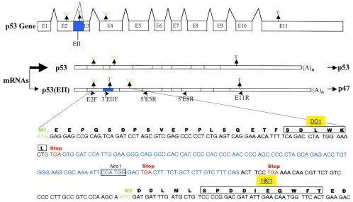
The development of cancer is a multistep process involving mutations in proto-oncogenes, tumor suppressor genes, and other genes which control cell proliferation, telomere stability, angiogenesis, and other complex traits. Despite this complexity, the cellular pathways controlled by the p53 tumor suppressor protein are compromised in most, if not all, cancers. In normal cells, p53 controls cell proliferation, senescence, and/or mediates apoptosis in response to stress, cell damage, or ectopic oncogene expression, properties which make p53 the prototype tumor suppressor gene. Defining the mechanisms of regulation of p53 activity in normal and tumor cells has therefore been a major priority in cell biology and cancer research. The present study reveals a novel and potent mechanism of p53 regulation originating through alternative splicing of the human p53 gene resulting in the expression of a novel p53 mRNA. This novel p53 mRNA encodes an N-terminally deleted isoform of p53 termed p47. As demonstrated within, p47 was able to effectively suppress p53-mediated transcriptional activity and impair p53-mediated growth suppression. It was possible to select for p53-null cells expressing p47 alone or coexpressing p53 in the presence of p47 but not cells expressing p53 alone. This showed that p47 itself does not suppress cell viability but could control p53-mediated growth suppression. Interestingly, p47 was monoubiquitinated in an Mdm2-independent manner, and this was associated with its export out of the nucleus. In the presence of p47, there was a reduction in Mdm2-mediated polyubiquitination and degradation of p53, and this was also associated with increased monoubiquitination and nuclear export of p53. The expression of p47 through alternative splicing of the p53 gene thus has a major influence over p53 activity at least in part through controlling p53 ubiquitination and cell localization.


Figures

FIG. 1.
FIG. 1.
Comparison of prototype p53 mRNA and p53(EII) mRNA. The top p53 gene diagram shows the novel EII exon (shown in blue) in the context of p53 exons 1 through 11. The two start codon methionines (M1 and M2) present in exons 2 and 4, respectively, are shown in green, and the stop codons (T) present in the novel EII exon and exon 11 are indicated in red. The middle diagram compares the p53(EII) mRNA to the prototype p53 mRNA, expressing p47 and p53, respectively, and the primers used to characterize these mRNAs, as detailed within. The bottom diagram shows the corresponding protein translation products derived from the p53(EII) mRNA and the locations of the epitopes for monoclonal antibodies DO1 and 1801.
FIG. 2.
FIG. 2.
Detection of p53(EII) mRNA in human cell lines and normal lymphocytes and analysis of its protein products. (A) (Left panel) Purified polyribosomal RNA from different human cell lines was subjected to reverse transcription-PCR analysis to identify the prototype p53 and p53(EII) mRNAs with the E2F/5′E5R and 3′EIIF/5′E5R primer pairs, respectively (see Fig. 1 for locations of primers). The resulting PCR products were digested with NcoI, which generates an 86-bp fragment from the p53(EII) PCR product but not from the prototype p53 PCR product (see Fig. 1 for location of the NcoI site in the EII exon). (Right panel) The same diagnostic reverse transcription-PCR followed by NcoI digestion as above was performed on total RNA isolated from normal human lymphocytes and different human cell lines. (B) Comparison of prototype p53 and p53(EII) mRNA levels. RNA was extracted from the indicated normal lymphocytes or established cell lines, and cDNA synthesis and kinetic quantitative PCR was carried out. To quantify p53 prototype plus EII exon-containing transcripts, primer set E2F/5′E5R was used (beaded lines). To quantify EII exon-containing transcripts, primer set 3′EIIF/5′E5R was used (solid lines). Data are representative of at least five independent experiments. (C) Western blot (WB) analysis of p53 and p47 in H1299 and 10(1) cells transfected with the indicated expression plasmids (see Fig. 1 for the locations of monoclonal antibody 1801 and DO1 epitopes). pCDNA3 represents the control plasmid-transfected cells. Note that the p53(EII) cDNA expressed both p53 and p47 proteins, as detected with monoclonal antibody 1801. (D) Western blot analysis of p53 and p47 in 10(1) cells transfected with p53(EII) cDNAs isolated from MCF-7 cells (lanes 3 and 4). For comparison, the first lane contains the p53(EII) cDNA derived from HeLa cells, and the second lane contains the p53 cDNA truncated in exons 1, 2, and EII as detailed in the text.
FIG. 3.
FIG. 3.
p47 inhibition of p53-mediated transcriptional transactivation and growth suppression. (A) (Top panel) p53-mediated transcriptional transactivation as determined by luciferase activity in H1299 and 10(1) cells cotransfected with a p53-responsive p21 promoter luciferase plasmid, the indicated p53 and p47 expression plasmids and a β-galactosidase expression plasmid to control for transfection efficiency. The p53-mediated transcriptional transactivation activity was expressed as relative light units (RLU) over pCDNA3, the control plasmid. (Bottom panel) The same cell lysates used for the luciferase assay were analyzed by Western blot with monoclonal antibody 1801 to detect p53 and p47 protein levels. This experiment was repeated four times with the same result. (B) p53-mediated suppression of colony formation is reversed by p47. H1299 cells were transfected with the empty vector (pCIN4) or with the indicated cDNAs as detailed in the text. The vector (pCIN4) used to express p53 and/or p47 contained the neomycin resistance gene, and therefore surviving colonies were selected for 2 weeks by addition of G418 to the culture medium, and drug-resistant colonies were counterstained with Giemsa. (C) Western blot analysis of H1299 cells stably expressing p47 or coexpressing p53 in the presence of p47. Cells as in panel B were pooled and subjected to Western blot analysis with monoclonal antibodies 1801 and DO1 as indicated. Note that no cell lines survived following selection for p53 expression alone with the pCIN4-p53 plasmid.
FIG. 4.
FIG. 4.
Association and oligomerization of p47 and p53. (A) Complexing of p53 with p47 in H1299 cells transfected with the p53(EII) cDNA. Control cells were also transfected with p53 or p47 expression plasmids. (Left panel) Transfected cell lysates were first subjected to immunoprecipitation with monoclonal antibody DO1, specific for p53, and the resultant precipitates were subjected to Western blot analysis with monoclonal antibody 1801 to detect both p53 and the coimmunoprecipitated p47. (Right panel) Western blot analysis with monoclonal antibody 1801 to determine p53 and p47 levels in the whole-cell lysates of the transfected cells used for the immunoprecipitation analysis. (B) Oligomerization analysis of p47 with p53. Cell lysates prepared from H1299 cells transfected with the indicated cDNAs were treated with 0, 0.01, or 0.1% glutaraldehyde for 5 min on ice. Treated lysates were resolved on a 4 to 15% gradient SDS-PAGE gel to differentiate monomers from oligomers. Western blot (WB) analysis was performed with monoclonal antibody 1801 (left panel), and the blot was stripped and reprobed with monoclonal antibody DO1 to detect p53-specific bands (right panel). Note that control p53-338 is a C-terminal mutant of p53 that is unable to oligomerize.
FIG. 5.
FIG. 5.
Control of p53 cell localization by p47 in transiently transfected and stable cell lines. (A) Percentage of cells with p53 and p47 localized predominantly in the nucleus or the cytoplasm in transiently transfected cells. The indicated expression plasmids were transfected into H1299 or 10(1) cells and the cell localization of p53 and p47 was determined by immunofluorescence (IF) microscopy with monoclonal antibody DO1 to detect p53 and monoclonal antibody 1801 to detect p47 in cells expressing only p47. Note that expression of p47 from the p53(EII) cDNA shifted the localization of p53 from the nucleus to the cytoplasm. The data are representative of three independent experiments. (B) Representative immunofluorescence images of 10(1) cells transiently transfected with the indicated expression cDNAs with monoclonal antibody DO1 specific for p53 and monoclonal antibody 1801 to detect p47. Note that, as expected, DO1 did not show any signal when used on p47-transfected cells because p47 lacks the DO1 N-terminal epitope. (C) Localization of p53 and p47 at day 1 and day 7 posttransfection with the indicated plasmids, where p53 was localized with monoclonal antibody DO1 and p47 was localized with monoclonal antibody 1801, as indicated. Note that on day 7, no surviving cells expressing p53 were detected. (D) Localization of p47 in stably transfected H1299 cells expressing p47 alone in untreated control cells and cells exposed to stress by transfecting empty plasmid pCDNA3 or treatment with adriamycin. (E) Localization of p53 in stably transfected H1299 cells coexpressing p53 and p47 in untreated control cells and cells exposed to stress by transfecting empty plasmid pCDNA3 or treatment with adriamycin.
FIG. 6.
FIG. 6.
Association and cell localization of p53 and p47 C-terminal oligomerization mutants. (A) Association analysis of p53 and p47 in H1299 cells transfected with the indicated plasmids expressing either wild-type or oligomerization-deficient p53, p53(EII), or p47 cDNAs. Oligomerization mutants p53-338, p53(EII)-338, and p47-338 lack the C terminus (amino acids 338 to 393), which includes the oligomerization domain. (Top panel) Transfected cell lysates were first subjected to immunoprecipitation with monoclonal antibody DO1 specific for p53, and the resultant immunoprecipitates were subjected to Western blot (WB) analysis with monoclonal antibody 1801 to detect both p53 and the coimmunoprecipitated p47. (Bottom panel) Western blot analysis with monoclonal antibody 1801 to determine p53 and p47 levels in the whole cell lysates of the transfected cells used for the immunoprecipitation analysis. (B) Cell localization of wild-type and oligomerization-deficient p53 and p47 proteins in H1299 cells 24 h posttransfection with the indicated plasmids. Cell localization of p53 and p47 was determined by immunofluorescence (IF) microscopy with monoclonal antibody DO1 to detect p53 and monoclonal antibody 1801 to detect p47 in cells expressing only p47. Note that in cells transfected with the p53(EII)-338 cDNA, in which p53 and p47 do not associate due to deletion of the oligomerization domain, p47 is no longer able to shift the localization of p53 to a more cytoplasmic distribution compared to cells transfected with the wild-type p53(EII) cDNA. The data are representative of three independent experiments.
FIG. 7.
FIG. 7.
Mdm2-mediated ubiquitination and degradation of p53 and p47. (A) H1299 cells were cotransfected with the indicated plasmids expressing p53, p53/p47 (p53[EII]), or p47 together with an HA-tagged ubiquitin expression plasmid and either control pCDNA3 plasmid or an Mdm2 expression plasmid as indicated. A β-galactosidase expression plasmid was also included to control for transfection efficiency. Cell lysates were prepared 24 h posttransfection and immunoprecipitated with either DO1 or 1801 followed by Western blot analysis with anti-HA antibody to detect ubiquitinated p53 and/or p47. Control lanes included cells coexpressing p53 or p47 with Mdm2 in the absence of HA-tagged ubiquitin (lanes 10 and 11) and cells expressing only HA-tagged ubiquitin (lane 12) revealed no ubiquitinated proteins, as expected. Note that in lane 1, no ubiquitinated p53 species were detected in the absence of Mdm2. Note also the predominance of monoubiquitinated species when p53 was coexpressed with p47 (lanes 3 and 4) or when p47 was expressed alone (lane 5) or in the presence of Mdm2 (lane 6). (B) Western blot analysis (monoclonal antibody 1801) of p53 and p47 protein levels in the whole-cell lysates (5% input) which were used to carry out the ubiquitination analysis shown in panel A. Note that the presence of p47 protects p53 from Mdm2-mediated degradation when comparing lanes 1 and 2 to lanes 3 and 4, and that Mdm2 was unable to mediate p47 degradation.

Similar articles

Cited by

References

    1. Balint, E., and K. Vousden. 2001. Activation and activities of the p53 tumor suppressor protein. Br. J. Cancer 85:1813-1823. - PMC - PubMed
    1. Boyd, S., Tsai, K., and Jacks, T. 2000. An intact HDM2 RING-finger domain is required for nuclear exclusion of p53. Nat. Cell Biol. 2:563-568. - PubMed
    1. Courtois, S., G. Verhaegh, S. North, M. G. Luciani, P. Lassus, U. Hibner, M. Oren, and P. Hainaut. 2002. DeltaN-p53, a natural isoform of p53 lacking the first transactivation domain, counters growth suppression by wild-type p53. Oncogene 21:6722-6728. - PubMed
    1. Donehower, L. 1996. The p53-deficient mouse: a model for basic and applied cancer studies. Semin. Cancer Biol. 7:269-278. - PubMed
    1. Freedman, D., and A. Levine. 1998. Nuclear export is required for degradation of endogenous p53 by Mdm2 and human papillomavirus E6. Mol. Cell. Biol. 18:7288-7293. - PMC - PubMed

Publication types

MeSH terms