. 2004 Sep;24(18):8037-47.
doi: 10.1128/MCB.24.18.8037-8047.2004.

Inactivation of Stat5 in mouse mammary epithelium during pregnancy reveals distinct functions in cell proliferation, survival, and differentiation

Affiliations

Inactivation of Stat5 in mouse mammary epithelium during pregnancy reveals distinct functions in cell proliferation, survival, and differentiation

Yongzhi Cui et al. Mol Cell Biol. 2004 Sep.

Abstract

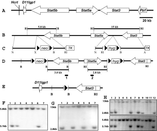
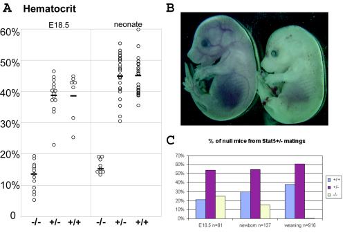
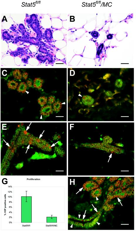
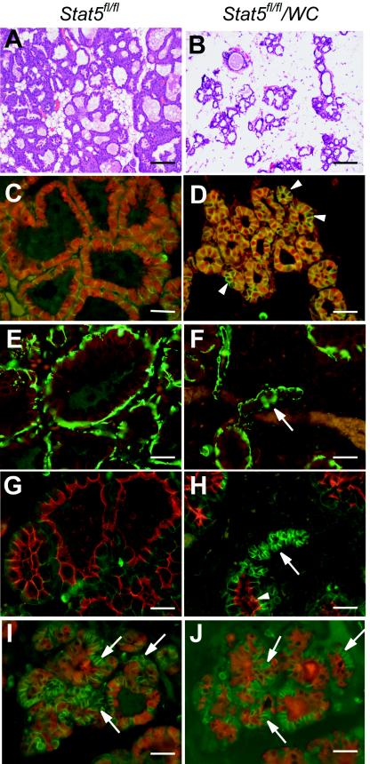
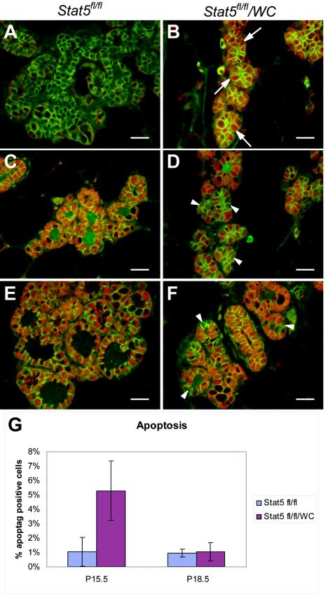
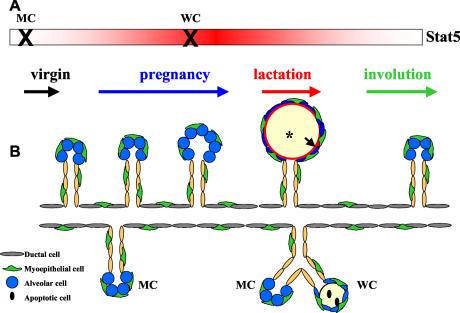
This study explored the functions of the signal transducers and activators of transcription 5a and 5b (referred to as Stat5 here) during different stages of mouse mammary gland development by using conditional gene inactivation. Mammary gland morphogenesis includes cell specification, proliferation and differentiation during pregnancy, cell survival and maintenance of differentiation throughout lactation, and cell death during involution. Stat5 is activated by prolactin, and its presence is mandatory for the proliferation and differentiation of mammary epithelium during pregnancy. To address the question of whether Stat5 is also necessary for the maintenance and survival of the differentiated epithelium, the two genes were deleted at different time points. The 110-kb Stat5 locus in the mouse was bracketed with loxP sites, and its deletion was accomplished by using two Cre-expressing transgenic lines. Loss of Stat5 prior to pregnancy prevented epithelial proliferation and differentiation. Deletion of Stat5 during pregnancy, after mammary epithelium had entered Stat5-mediated differentiation, resulted in premature cell death, indicating that at this stage epithelial cell proliferation, differentiation, and survival require Stat5.


Figures

FIG. 1.
FIG. 1.
Introduction of loxP sites into the Stat5a/b locus. (A) The BAC clone spans approximately 210 kb and encompasses five known genes and one new gene. The Stat5a gene is 30 kb, Stat5b is more than 54 kb, and Stat3 is more than 37 kb. The Hcrt and D11lgp1 genes are downstream of the Stat5b gene, and the Ptrf gene is upstream of the Stat3 gene. The gene sizes and intergenic sequences are drawn to scale. (B) Restriction map of the wild-type allele. B, BamHI; RI, EcoRI. (C) The Stat5b-targeting construct contains a 7.2-kb genomic sequence with a pLoxpneo cassette, and the Stat5a-targeting vector contains an 8.6-kb genomic sequence with a pLoxphyg cassette. Neomycin (neo), hygromycin (hyg), and thymidine kinase (TK) are positive and negative selection cassettes, respectively. Prior to electroporation, each targeting vector was linearized at the unique NotI site (N) indicated. X, XbaI; A, Asp718. (D) Restriction map of the targeted allele. Two loxP sites and the neomycin cassette were introduced downstream of Stat5b, and two loxP sites with the hygromycin cassette were inserted downstream of Stat5a. The indicated probes were used to assess recombination events. (E) Structure of the deleted Stat5a/b allele. D11lgp1 and Stat3 are next to each other, with one loxP site between them. (F) Southern blot analysis of homologous recombination in ES cells electroporated with the Stat5b-targeting vector. The BamHI (lanes 1, 2, and 6) restriction fragment size changes from 5.8 to 3.0 kb, as seen in the targeted cells with the 32P-labeled 500-bp external probe 1. (G) ES cell clone 1 was chosen to be electroporated with the Stat5a-targeting vector. The EcoRI (lanes 1, 3, 5, and 7) restriction fragment size changes from 15 to 2.8 kb, as seen in the targeted cells with the 32P-labeled 500-bp external probe 2. (H) Southern blot analysis of tail DNA from chimeric males (lanes 1 and 2) and their agouti pups (lanes 3 to 12). Both Stat5a- and Stat5b-targeted bands were detected in the male chimeras (lanes 1 and 2) and in four agouti pups (lanes 6, 8, 9, and 12).
FIG. 2.
FIG. 2.
Stat5-null embryos are perinatal lethal. (A) Hematocrits of Stat5−/− (n = 13 and 9), Stat5+/− (n = 15 and 28), or Stat5+/+ (n = 7 and 25) E18.5 fetuses and newborn littermates. Differences between Stat5+/+ and Stat5−/− animals were highly significant (P < 0.0001 and P < 10−18 by the two-tailed Student t test). There was no difference between wild-type and hemizygous animals (P = 0.913 and P = 0.676). (B) An E14.5 Stat5−/− embryo (right) is paler and smaller than its wild-type littermate (left). (C) The Mendelian ratio (25%; n = 81) was observed in E18.5 Stat5−/− fetuses; it dropped to 15% (n = 137) in the neonates, and only 0.5% (n = 916) were Stat5 null at weaning.
FIG. 3.
FIG. 3.
Reduced proliferation of epithelial cells in the absence of Stat5. Stat5fl/fl and Stat5fl/fl/MC virgin mice (12 weeks) were injected with estrogen and progesterone. Forty-eight hours after treatment, mammary glands were harvested and fixed. (A and B) H&E-stained sections showed sparse branching in the Stat5fl/fl/MC glands compared to Stat5fl/fl littermate controls after acute estrogen-progesterone treatment. (C and D) Most mammary epithelial cells in estrogen-progesterone-treated Stat5fl/fl/MC virgin mice were devoid of Stat5 as demonstrated by immunofluorescence staining with Stat5a (red) and E-cadherin (green) antibodies. Occasional Stat5-expressing cells were observed (D) (arrowhead) in Stat5fl/fl/MC epithelia due to the mosaic expression of Cre recombinase. (E and F) Immunofluorescence staining showed abundant H3P (a proliferation marker, in red) in estrogen-progesterone-treated Stat5fl/fl mammary tissues (E) (arrows), it but was found only in few cells in Stat5fl/fl/MC epithelia(F) (arrow). E-cadherin (green) was used as a cell membrane stain. (G) H3P staining demonstrated that 2.1% ± 0.67% (mean ± SEM) of mammary cells in Stat5fl/fl/MC mice were positive for H3P, while 10.2% ± 2.2% of cells were stained in control mice. The difference was statistically significant (P < 0.0001 by the two-tailed Student t test). (H) Immunofluorescence staining showed Cre recombinase (red, arrows) expression in the mammary tissue of a Stat5fl/fl/MC virgin animal. E-cadherin is in green. MMTV-Cre expression was mosaic in ductal mammary epithelium (arrows and arrowheads), and only a few cells expressed Cre recombinase (arrows). Bars, 50 μm.
FIG.4.
FIG.4.
Stat5 is required for functional differentiation of the mammary epithelium. Inguinal mammary glands were collected from Stat5fl/fl (A, C, E, and G) and Stat5fl/fl/WC (B, D, F, H, I, and J) mice at lactation. (A and B) H&E staining showed sparser alveoli in Stat5fl/fl/WC mammary gland at lactation than in the Stat5fl/fl littermate. (C to J) Mammary tissues were harvested at parturition and stained with antibodies that characterize the differentiation state of epithelial cells. Uniform nuclear Stat5 (red) staining was seen in the Stat5fl/fl gland (C). However, a conspicuous nonuniform staining of Stat5 was observed in most alveoli in Stat5fl/fl/WC tissue (D) (arrowhead). NKCC1 (red) expression was undetectable in Stat5fl/fl/WC and Stat5fl/fl epithelia during lactation (E and F) (arrow); smooth muscle actin (green) stained the myoepithelial cells. Alveoli in Stat5fl/fl/WC displayed heterogeneous Npt2b staining (red) at parturition (H) (arrow). While this marker for secretory activity was seen on the apical membrane in expanded alveoli, it was absent in alveoli with a small lumen (G and H) (arrowhead). E-cadherin (green) was used to stain the cell membrane. (I and J) Serial sections from a Stat5fl/fl/WC gland stained with Stat5 (red) (I) and WAP (red) (J); E-cadherin (green) was applied in both sections. Note that cells without Stat5a (I) (arrows) do not express WAP at parturition (J) (arrows). Bars, 250 μm (A and B) and 50 μm (C to J).
FIG.5.
FIG.5.
Stat5 is required for survival of the differentiated mammary epithelium. (A to D) Inguinal mammary glands were collected from Stat5fl/fl (A and C) and Stat5fl/fl/WC (B and D) mice at p15.5. (E and F) Mammary tissues harvested at p18.5. (A and B) Immunofluorescence staining of Cre recombinase (red) and E-cadherin (green) showed a mosaic pattern of WAP-Cre expression in p15.5 Stat5fl/fl/WC mammary tissue (B) (arrows), and no WAP-Cre was detected in Stat5fl/fl mice (A). (C and D) Uniform Stat5a (red) expression was observed in Stat5fl/fl epithelium at p15.5, while about one-half of the epithelial cells had lost Stat5a expression in Stat5fl/fl/WC mice (D) (arrowheads). E-cadherin (green) was used to stain cell membranes. (E and F) At p18.5 only a few mammary epithelial cells in Stat5fl/fl/WC mice were negative for Stat5a (F) (arrowheads). E-cadherin was stained as green. (G) TUNEL assays were performed with the inguinal mammary glands at p15.5 and p18.5 from three Stat5fl/fl and six Stat5fl/fl/WC mice. At p15.5, 5.29% ± 2.07% (mean ± SEM) TUNEL-positive cells were observed in Stat5fl/fl/WC epithelium, compared with 1.02% ± 1.0% in Stat5fl/fl tissue. The difference was highly significant (P = 0.0046 by the two-tailed Student t test). In contrast, at p18.5 the number of apoptotic cells decreased to 1.03% ± 0.64% in the Stat5fl/fl/WC mammary gland, which was comparable to the percentage of positive cells (0.97% ± 0.27%) in Stat5fl/fl mice (P = 0.798).
FIG. 6.
FIG. 6.
Stat5 controls proliferation, differentiation, and survival of mammary alveolar epithelium. Wild-type mammary epithelium differentiates into functional alveoli during pregnancy. When Stat5 deletion occurs prior to pregnancy, the mammary epithelium loses the ability to respond to proliferative signals at the onset of pregnancy; it retains ductal characteristics and fails to acquire a marker indicative of secretory function. When the loss of Stat5 occurs late in pregnancy after mammary epithelia have entered differentiation, differentiation is stalled and premature cell death takes place. (A) The rectangle indicates that Stat5 is expressed throughout mammary gland development, and the red gradient reflects Stat5 activity during pregnancy and lactation. The X refers to the time when Stat5 is inactivated. MC, Stat5 deletion occurs before puberty in Stat5fl/fl/MC mouse. WC, Stat5 deletion takes place during late pregnancy. (B) Schematic presentation of mammary epithelial development in the presence and absence of Stat5. Ducts and alveoli in the upper part depict mammary epithelial development in wild-type mice. Ducts and alveoli in the lower part illustrate the abnormal mammary epithelial development in Stat5fl/fl/MC or Stat5fl/fl/WC mice during pregnancy and lactation. *, milk secretion in the alveoli. The arrow points to the Npt2b localization in the functional alveoli.

References

    1. Acosta, J. J., R. M. Munoz, L. Gonzalez, A. Subtil-Rodriguez, M. A. Dominguez-Caceres, J. M. Garcia-Martinez, A. Calcabrini, I. Lazaro-Trueba, and J. Martin-Perez. 2003. Src mediates prolactin-dependent proliferation of T47D and MCF7 cells via the activation of focal adhesion kinase/Erk1/2 and phosphatidylinositol 3-kinase pathways. Mol. Endocrinol. 17:2268-2282. - PubMed
    1. Bunting, K. D., H. L. Bradley, T. S. Hawley, R. Moriggl, B. P. Sorrentino, and J. N. Ihle. 2002. Reduced lymphomyeloid repopulating activity from adult bone marrow and fetal liver of mice lacking expression of STAT5. Blood 99:479-487. - PubMed
    1. Burdon, T., L. Sankaran, R. J. Wall, M. Spencer, and L. Hennighausen. 1991. Expression of a whey acidic protein transgene during mammary development. Evidence for different mechanisms of regulation during pregnancy and lactation. J. Biol. Chem. 266:6909-6914. - PubMed
    1. Cui, Y., M. Li, K. D. Walton, K. Sun, J. A. Hanover, P. A. Furth, and L. Hennighausen. 2001. The Stat3/5 locus encodes novel endoplasmic reticulum and helicase-like proteins that are preferentially expressed in normal and neoplastic mammary tissue. Genomics 78:129-134. - PubMed
    1. Deng, C., A. Wynshaw-Boris, F. Zhou, A. Kuo, and P. Leder. 1996. Fibroblast growth factor receptor 3 is a negative regulator of bone growth. Cell 84:911-921. - PubMed

MeSH terms

LinkOut - more resources