. 2004 Sep;24(18):8069-79.
doi: 10.1128/MCB.24.18.8069-8079.2004.

Sgt1 associates with Hsp90: an initial step of assembly of the core kinetochore complex

Affiliations

Sgt1 associates with Hsp90: an initial step of assembly of the core kinetochore complex

Parmil K Bansal et al. Mol Cell Biol. 2004 Sep.

Abstract

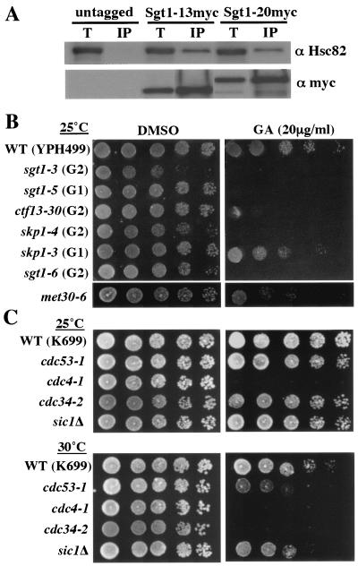
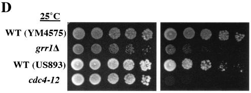
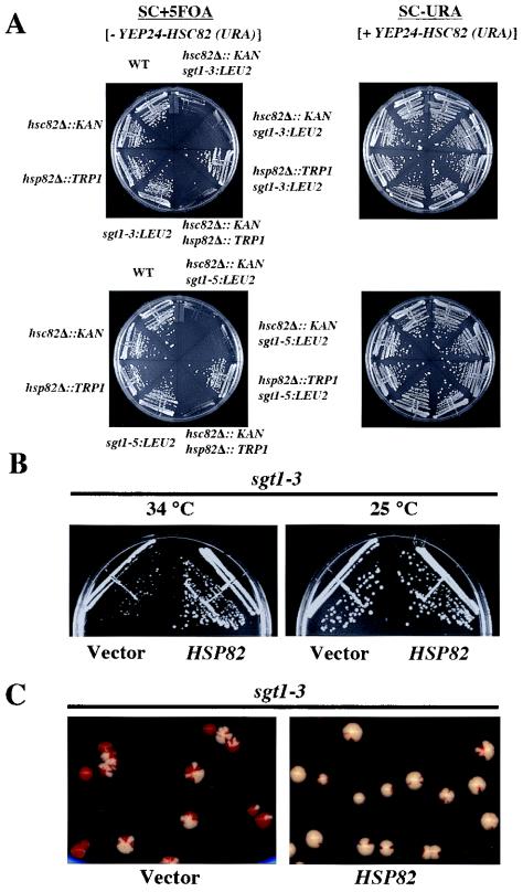
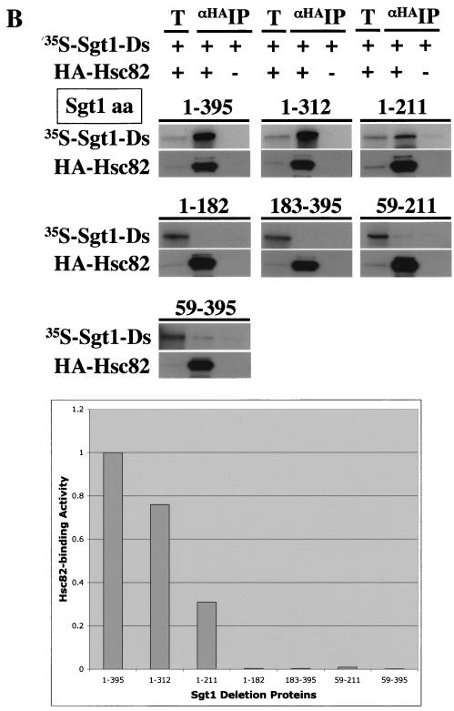
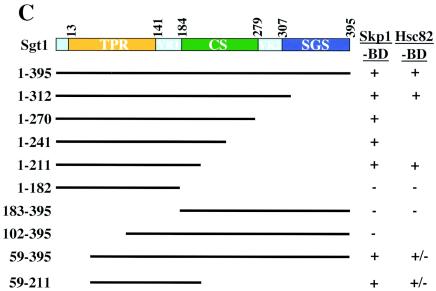
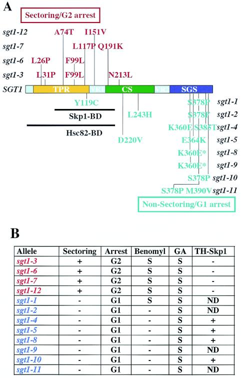
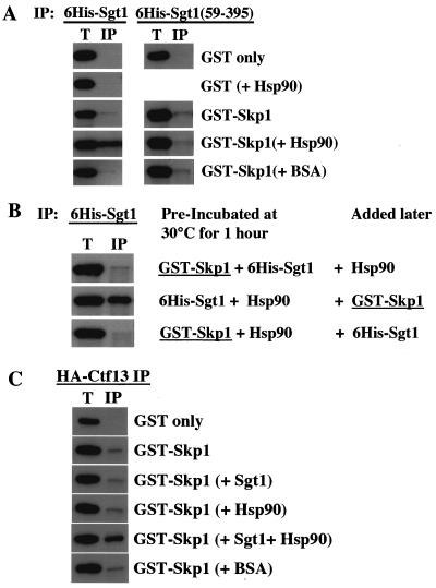
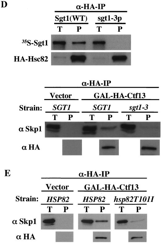
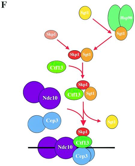
The kinetochore, which consists of DNA sequence elements and structural proteins, is essential for high-fidelity chromosome transmission during cell division. In budding yeast, Sgt1, together with Skp1, is required for assembly of the core kinetochore complex (CBF3) via Ctf13 activation. Formation of the active Ctf13-Skp1 complex also requires Hsp90, a molecular chaperone. We have found that Sgt1 interacts with Hsp90 in yeast. We also have determined that Skp1 and Hsc82 (a yeast Hsp90 protein) bind to the N-terminal region of Sgt1 that contains tetratricopeptide repeat motifs. Results of sequence and phenotypic analyses of sgt1 mutants strongly suggest that the N-terminal region containing the Hsc82-binding and Skp1-binding domains of Sgt1 is important for the kinetochore function of Sgt1. We found that Hsp90's binding to Sgt1 stimulates the binding of Sgt1 to Skp1 and that Sgt1 and Hsp90 stimulate the binding of Skp1 to Ctf13, the F-box core kinetochore protein. Our results strongly suggest that Sgt1 and Hsp90 function in assembling CBF3 by activating Skp1 and Ctf13.


Figures

FIG. 1.
FIG. 1.
Sgt1 associates with Hsp90 in yeast. (A) Proteins from strains expressing untagged Sgt1 or myc-tagged Sgt1 (Sgt1-13myc and Sgt1-20myc) were immunoprecipitated with anti-myc antibody and then immunoblotted with anti-Hsc82 antibody. T, total lysate; IP, immunoprecipitate. (B) The sgt1 mutants and SCF mutants are sensitive to geldanamycin. The indicated strains were grown on yeast extract-peptone-dextrose plates containing 20 μg of geldanamycin per ml (GA) or dimethyl sulfoxide (DMSO) only. The numbers of cells that were spotted onto each plate (left to right) were approximately 5 × 10⁴, 1 × 10⁴, 2 × 10³, 4 × 10², and 8 × 101. The plates were incubated at the indicated permissive temperatures for 3 days. (B) Strains in the YPH499 background. (C) Strains in the K699 background. (D) Strains in the YM4575 or US893 background. WT, wild type.
FIG. 1.
FIG. 1.
Sgt1 associates with Hsp90 in yeast. (A) Proteins from strains expressing untagged Sgt1 or myc-tagged Sgt1 (Sgt1-13myc and Sgt1-20myc) were immunoprecipitated with anti-myc antibody and then immunoblotted with anti-Hsc82 antibody. T, total lysate; IP, immunoprecipitate. (B) The sgt1 mutants and SCF mutants are sensitive to geldanamycin. The indicated strains were grown on yeast extract-peptone-dextrose plates containing 20 μg of geldanamycin per ml (GA) or dimethyl sulfoxide (DMSO) only. The numbers of cells that were spotted onto each plate (left to right) were approximately 5 × 10⁴, 1 × 10⁴, 2 × 10³, 4 × 10², and 8 × 101. The plates were incubated at the indicated permissive temperatures for 3 days. (B) Strains in the YPH499 background. (C) Strains in the K699 background. (D) Strains in the YM4575 or US893 background. WT, wild type.
FIG. 2.
FIG. 2.
(A) The sgt1 and hsc82 mutations exert synthetic lethality. The indicated single and double mutants carrying YEP24-HSC82 were streaked onto plates containing Sc+5-FOA and Sc−Ura, respectively. The plates were incubated at 25°C for 2 days. (B) Hsp82 overexpression suppressed the temperature sensitivity of sgt1-3. The sgt1-3 cells containing p413-GPD-Hsp82 (GPD promoter/CEN/HIS3) and the vector alone were streaked onto Sc-His plates and incubated at the indicated temperatures for 3 days. (C) Hsp82 overexpression suppressed the chromosome missegregation phenotype of sgt1-3 cells. Shown are the results of a colony color-sectoring assay to analyze sgt1-3 cells that contained the vector alone and that contained p413-GPD-Hsp82. WT, wild type.
FIG. 3.
FIG. 3.
Skp1-binding and Hsc82-binding domains of Sgt1. (A, top) The indicated proteins were expressed and labeled with [35S]methionine in the in vitro translation system with rabbit reticulocyte lysates, and the radiolabeled proteins were mixed and incubated for 1 h at 30°C with an extract containing HA-tagged Skp1 that was not radiolabeled (20, 22). HA-tagged Skp1 was immunoprecipitated by using anti-HA-Sepharose, the immunoprecipitates were eluted and subjected to SDS-PAGE, and the radioactive bands were identified by autoradiography. HA-tagged Skp1 (HA-Skp1) was detected by immunoblotting with anti-HA antibody. T, total lysates (6% of the starting material); IP, immunoprecipitate; Ds, deletion proteins. The numbers above the lanes indicate the positions of the N- and C-terminal amino acids (aa) in each Sgt1 deletion protein. (A, bottom) Relative Skp1-binding activities of Sgt1 mutant proteins. Binding activity was defined as the ratio of the amount of coprecipitated protein to the amount of input protein. The binding activity of Sgt1 (wild type) was defined as 1, and the binding activities of the mutants were normalized to that value. (B, top) Essentially the same experiments as in panel A but with HA-tagged Hsc82 in place of HA-tagged Skp1. HA-tagged Hsc82 was expressed in the in vitro translation system but not radiolabeled. HA-tagged Hsc82 was detected by immunoblotting with anti-HA antibody. The numbers in parentheses indicate the positions of the N- and C-terminal amino acids in each Sgt1 deletion protein. (B, bottom) Relative Hsc82-binding activities of Sgt1 mutant proteins. Binding activity was calculated as described for panel A. (C) Illustration of the Sgt1 deletion proteins used in the Skp1- and Hsc82-binding assays and the results of these assays. The results of the Skp1-binding domain (Skp1-BD) and the Hsc82-binding domain (Hsc82-BD) analyses are summarized. Plus signs indicate significant binding activity. Minus signs indicate no significant binding activity. Plus-minus signs indicate weak binding activity.
FIG. 3.
FIG. 3.
Skp1-binding and Hsc82-binding domains of Sgt1. (A, top) The indicated proteins were expressed and labeled with [35S]methionine in the in vitro translation system with rabbit reticulocyte lysates, and the radiolabeled proteins were mixed and incubated for 1 h at 30°C with an extract containing HA-tagged Skp1 that was not radiolabeled (20, 22). HA-tagged Skp1 was immunoprecipitated by using anti-HA-Sepharose, the immunoprecipitates were eluted and subjected to SDS-PAGE, and the radioactive bands were identified by autoradiography. HA-tagged Skp1 (HA-Skp1) was detected by immunoblotting with anti-HA antibody. T, total lysates (6% of the starting material); IP, immunoprecipitate; Ds, deletion proteins. The numbers above the lanes indicate the positions of the N- and C-terminal amino acids (aa) in each Sgt1 deletion protein. (A, bottom) Relative Skp1-binding activities of Sgt1 mutant proteins. Binding activity was defined as the ratio of the amount of coprecipitated protein to the amount of input protein. The binding activity of Sgt1 (wild type) was defined as 1, and the binding activities of the mutants were normalized to that value. (B, top) Essentially the same experiments as in panel A but with HA-tagged Hsc82 in place of HA-tagged Skp1. HA-tagged Hsc82 was expressed in the in vitro translation system but not radiolabeled. HA-tagged Hsc82 was detected by immunoblotting with anti-HA antibody. The numbers in parentheses indicate the positions of the N- and C-terminal amino acids in each Sgt1 deletion protein. (B, bottom) Relative Hsc82-binding activities of Sgt1 mutant proteins. Binding activity was calculated as described for panel A. (C) Illustration of the Sgt1 deletion proteins used in the Skp1- and Hsc82-binding assays and the results of these assays. The results of the Skp1-binding domain (Skp1-BD) and the Hsc82-binding domain (Hsc82-BD) analyses are summarized. Plus signs indicate significant binding activity. Minus signs indicate no significant binding activity. Plus-minus signs indicate weak binding activity.
FIG. 4.
FIG. 4.
Mutations in temperature-sensitive sgt1 mutants. (A) The mutations in sgt1 G2 alleles are in red; the mutations in sgt1 G1 alleles are in blue. “Arrest” indicates the cell cycle stage at which the mutant cells accumulate when incubated at the nonpermissive temperature (unpublished data). Skp1-BD, Skp1-binding domain of Sgt1; Hsc82-BD, Hsc82-binding domain of Sgt1. The mutants indicated by the asterisk in panel A differ: sgt1-8 has two additional silent mutations at nucleotide positions 403 and 864. (Table 3 contains details of the sgt1 mutations.) (B) Summary of the phenotypes of sgt1 mutants. Plus signs in the sectoring column indicate that the mutant cell showed a chromosome missegregation defect that was shown by a colony color-sectoring assay. Minus signs in the sectoring column indicate that the cells did not show substantial chromosome missegregation. The letter S indicates sensitivity to benomyl or geldanamycin (GA); a minus sign indicates resistance. Because the sgt1-10 mutant, which has only the S378P mutation, is not sensitive to benomyl, we believe that the Y119C mutation is probably responsible for the benomyl sensitivity of the sgt1-1 mutant, which has two mutations (Y119C and S378P). Also shown are results of the two-hybrid assay (TH-Skp1) to assess the ability of sgt1 mutant proteins to bind to Skp1 (unpublished data). A plus sign in the TH-Skp1 column indicates that the mutant protein bound to Skp1 in the two-hybrid system. A minus sign indicates that the mutant protein did not do so. ND, not done.
FIG. 5.
FIG. 5.
Sgt1, together with Hsp90, stimulates Skp1-Ctf13 binding. (A) Hsp90 requires the N-terminal region of Sgt1 to promote its interaction with Skp1. Approximately 200 ng of each indicated protein was used for immunoprecipitation (IP). After the samples were incubated at 30°C for 1 h, His6-tagged Sgt1 or His6-tagged Sgt1(59-395) was immunoprecipitated with anti-His antibody conjugated to Sepharose. GST-Skp1 and GST proteins were detected by immunoblotting. The bands in the top two panels are GST protein only, and the bands in the bottom three panels are the GST-Skp1 fusion protein. The names of proteins in parentheses indicate that these proteins were present in the reaction mixture but are not shown. BSA, bovine serum albumin. (B) Experiment similar to that described for panel A but with preincubation of two of the three indicated proteins at 30°C for 1 h and addition of the other later (immediately before immunoprecipitation). (C) Sgt1 and Hsp90 stimulate the binding of Ctf13 to Skp1. This experiment was conducted as described for panel A, except that HA-tagged Ctf13 was precipitated with anti-HA antibody conjugated to Sepharose. The bands in the top panel are GST protein only, and the bands in the bottom five panels are the GST-Skp1 protein. (D, top) sgt1-3 protein (sgt1-3p) does not bind to Hsc82 in vitro. Wild-type (WT) Sgt1 protein and sgt1-3 mutant protein were expressed and labeled with [35S]methionine in the in vitro translation system, and the radiolabeled proteins were mixed and incubated for 1 h at 30°C with an extract containing HA-tagged Hsc82 (HA-Hsc82) that was not radiolabeled. HA-Hsc82 was immunoprecipitated by using anti-HA-Sepharose, the immunoprecipitates were eluted and subjected to SDS-PAGE, and the radioactive bands were identified by autoradiography. HA-Hsc82 was detected by immunoblotting with anti-HA antibody. T, total lysates (6% of the starting material); P, immunoprecipitate. (D, bottom) The Skp1-Ctf13 association was reduced in sgt1-3 cells. HA-tagged Ctf13 expression was induced by addition of galactose; 1 h later, the temperature was shifted to 37°C and the cells were incubated for 90 min. HA-tagged Ctf13 was immunoprecipitated by using anti-HA antibody conjugated to Sepharose. Skp1 was detected by Western blot analysis with anti-Skp1 antibody. (E) The Skp1-Ctf13 association was reduced in hsp82T101I cells. HA-tagged Ctf13 expression was induced by addition of galactose; 1 h later, the temperature was shifted to 37°C and the cells were incubated for 90 min. HA-tagged Ctf13 was immunoprecipitated by using anti-HA antibody conjugated to Sepharose. Skp1 was detected by Western blot analysis with anti-Skp1 antibody. (F) Model of CBF3 assembly. Once Sgt1 forms a complex containing Hsp90, Sgt1 is activated to bind to Skp1. By binding to Sgt1, Skp1 is activated to bind to Ctf13 (34).
FIG. 5.
FIG. 5.
Sgt1, together with Hsp90, stimulates Skp1-Ctf13 binding. (A) Hsp90 requires the N-terminal region of Sgt1 to promote its interaction with Skp1. Approximately 200 ng of each indicated protein was used for immunoprecipitation (IP). After the samples were incubated at 30°C for 1 h, His6-tagged Sgt1 or His6-tagged Sgt1(59-395) was immunoprecipitated with anti-His antibody conjugated to Sepharose. GST-Skp1 and GST proteins were detected by immunoblotting. The bands in the top two panels are GST protein only, and the bands in the bottom three panels are the GST-Skp1 fusion protein. The names of proteins in parentheses indicate that these proteins were present in the reaction mixture but are not shown. BSA, bovine serum albumin. (B) Experiment similar to that described for panel A but with preincubation of two of the three indicated proteins at 30°C for 1 h and addition of the other later (immediately before immunoprecipitation). (C) Sgt1 and Hsp90 stimulate the binding of Ctf13 to Skp1. This experiment was conducted as described for panel A, except that HA-tagged Ctf13 was precipitated with anti-HA antibody conjugated to Sepharose. The bands in the top panel are GST protein only, and the bands in the bottom five panels are the GST-Skp1 protein. (D, top) sgt1-3 protein (sgt1-3p) does not bind to Hsc82 in vitro. Wild-type (WT) Sgt1 protein and sgt1-3 mutant protein were expressed and labeled with [35S]methionine in the in vitro translation system, and the radiolabeled proteins were mixed and incubated for 1 h at 30°C with an extract containing HA-tagged Hsc82 (HA-Hsc82) that was not radiolabeled. HA-Hsc82 was immunoprecipitated by using anti-HA-Sepharose, the immunoprecipitates were eluted and subjected to SDS-PAGE, and the radioactive bands were identified by autoradiography. HA-Hsc82 was detected by immunoblotting with anti-HA antibody. T, total lysates (6% of the starting material); P, immunoprecipitate. (D, bottom) The Skp1-Ctf13 association was reduced in sgt1-3 cells. HA-tagged Ctf13 expression was induced by addition of galactose; 1 h later, the temperature was shifted to 37°C and the cells were incubated for 90 min. HA-tagged Ctf13 was immunoprecipitated by using anti-HA antibody conjugated to Sepharose. Skp1 was detected by Western blot analysis with anti-Skp1 antibody. (E) The Skp1-Ctf13 association was reduced in hsp82T101I cells. HA-tagged Ctf13 expression was induced by addition of galactose; 1 h later, the temperature was shifted to 37°C and the cells were incubated for 90 min. HA-tagged Ctf13 was immunoprecipitated by using anti-HA antibody conjugated to Sepharose. Skp1 was detected by Western blot analysis with anti-Skp1 antibody. (F) Model of CBF3 assembly. Once Sgt1 forms a complex containing Hsp90, Sgt1 is activated to bind to Skp1. By binding to Sgt1, Skp1 is activated to bind to Ctf13 (34).

Similar articles

Cited by

References

    1. Austin, M. J., P. Muskett, K. Kahn, B. J. Feys, J. D. Jones, and J. E. Parker. 2002. Regulatory role of SGT1 in early R gene-mediated plant defenses. Science 295:2077-2080. - PubMed
    1. Azevedo, C., A. Sadanandom, K. Kitagawa, A. Freialdenhoven, K. Shirasu, and P. Schulze-Lefert. 2002. The RAR1 interactor SGT1, an essential component of R gene-triggered disease resistance. Science 295:2073-2076. - PubMed
    1. Borkovich, K. A., F. W. Farrelly, D. B. Finkelstein, J. Taulien, and S. Lindquist. 1989. hsp82 is an essential protein that is required in higher concentrations for growth of cells at higher temperatures. Mol. Cell. Biol. 9:3919-3930. - PMC - PubMed
    1. Connelly, C., and P. Hieter. 1996. Budding yeast SKP1 encodes an evolutionarily conserved kinetochore protein required for cell cycle progression. Cell 86:275-285. - PMC - PubMed
    1. Doheny, K. F., P. K. Sorger, A. A. Hyman, S. Tugendreich, F. Spencer, and P. Hieter. 1993. Identification of essential components of the S. cerevisiae kinetochore. Cell 73:761-774. - PMC - PubMed

Publication types

MeSH terms