Comparative Study
. 2004 Sep;24(18):8276-87.
doi: 10.1128/MCB.24.18.8276-8287.2004.

Yeast Asc1p and mammalian RACK1 are functionally orthologous core 40S ribosomal proteins that repress gene expression

Affiliations
Comparative Study

Yeast Asc1p and mammalian RACK1 are functionally orthologous core 40S ribosomal proteins that repress gene expression

Vincent R Gerbasi et al. Mol Cell Biol. 2004 Sep.

Abstract

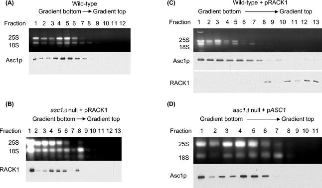
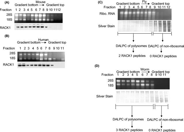
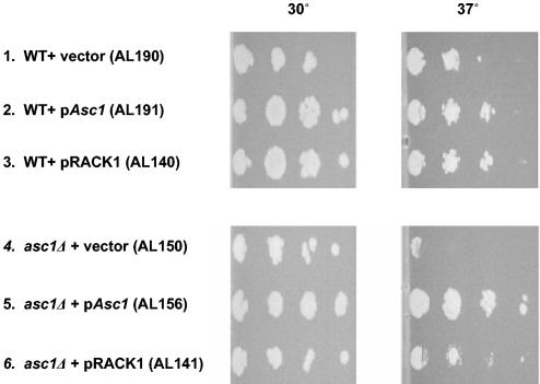
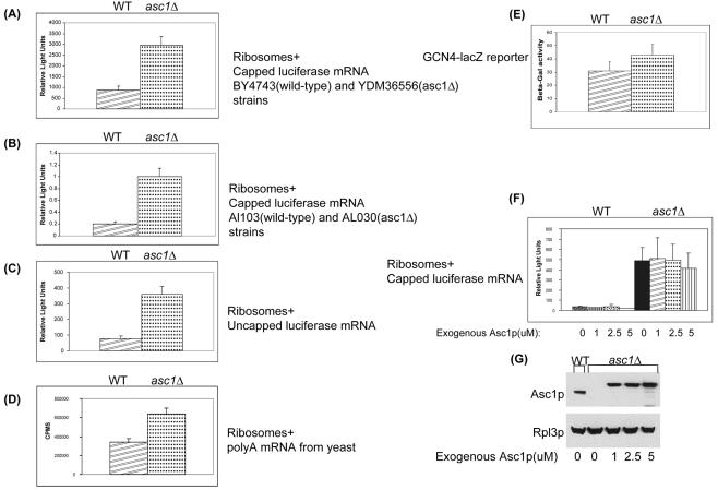
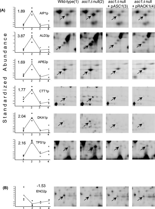
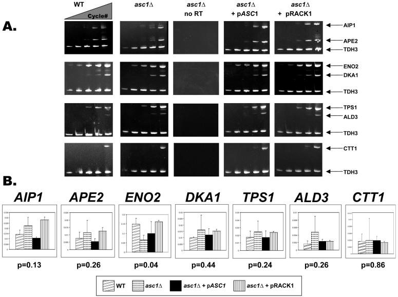
Translation of mRNA into protein is a fundamental step in eukaryotic gene expression requiring the large (60S) and small (40S) ribosome subunits and associated proteins. By modern proteomic approaches, we previously identified a novel 40S-associated protein named Asc1p in budding yeast and RACK1 in mammals. The goals of this study were to establish Asc1p or RACK1 as a core conserved eukaryotic ribosomal protein and to determine the role of Asc1p or RACK1 in translational control. We provide biochemical, evolutionary, genetic, and functional evidence showing that Asc1p or RACK1 is indeed a conserved core component of the eukaryotic ribosome. We also show that purified Asc1p-deficient ribosomes have increased translational activity compared to that of wild-type yeast ribosomes. Further, we demonstrate that asc1Delta null strains have increased levels of specific proteins in vivo and that this molecular phenotype is complemented by either Asc1p or RACK1. Our data suggest that one of Asc1p's or RACK1's functions is to repress gene expression.


Figures

FIG. 1.
FIG. 1.
Polysome profiles showing that Asc1p and RACK1 are biochemically orthologous ribosomal proteins. (A) Polysome profile showing that yeast Asc1p localizes to the polysome fractions and is absent in the nonribosomal fractions. Cell lysate from yeast wild-type strain AL190 was fractionated by sucrose gradient centrifugation, and fractions were collected. An aliquot of each fraction was analyzed by agarose gel electrophoresis to identify fractions with 25S and 18S rRNAs. Western analysis of the fractions with anti-Asc1p antibodies shows that Asc1p is in the ribosome fractions (lanes 1 to 8) and is absent from the nonribosomal fractions (lanes 9 to 12). (B) Polysome profile showing that RACK1 expressed in a yeast asc1Δ null strain localizes to the ribosomal fractions and is absent from the nonribosomal fractions. Cell lysate from yeast strain AL141 was fractionated by sucrose gradient centrifugation and analyzed as described for panel A except that anti-RACK1 antibody was used for Western analysis. (C) Polysome profile showing exclusion of RACK1 from ribosomal fractions in a wild-type yeast strain. Cell lysate from yeast strain AL140 was fractionated by sucrose gradient centrifugation and analyzed as described for panel A except that anti-RACK1 and anti-Asc1p antibodies were used in separate Western blot assays. (D) Polysome profile of an asc1Δ null strain complemented by expression of ASC1. Cell lysate from yeast strain AL156 was fractionated by sucrose gradient centrifugation and analyzed as described for panel A.
FIG. 2.
FIG. 2.
Polysome profiling of RACK1 protein showing its polysomal localization in four eukaryotic species. (A) Mouse polysome profile showing that RACK1 localizes to the polysome fractions and is absent from the nonribosomal fractions. A cell lysate from mouse NT2 cells was fractionated by sucrose gradient centrifugation, and fractions were collected. An aliquot of each fraction was analyzed by agarose gel electrophoresis to show fractions with 28S and 18S rRNAs. Western analysis of the fractions with anti-RACK1 antibodies shows the distribution of RACK1. (B) Human polysome profile showing that RACK1 localizes to the polysome fractions and is absent from the nonribosomal fractions. A cell lysate from human HEK293 cells was fractionated by sucrose gradient centrifugation and analyzed as described for panel A. (C) DALPC analysis of the Drosophila polysome profile shows that the putative RACK1 ortholog (NP_477269) localizes to the polysome fractions and is absent from the nonribosomal fractions. Each fraction was analyzed by sodium dodecyl sulfate-polyacrylamide gel electrophoresis to visualize the amount of protein in each fraction. Polysome and nonribosomal fractions were separately pooled and digested with trypsin, and proteins were identified by the DALPC-MS approach (34, 47, 63). The Drosophila 28S and 18S rRNAs typically comigrate as a doublet (27). Ribo., ribosomal. (D) DALPC analysis of a nematode polysome profile shows that the putative worm RACK1 ortholog (NP_501859) localizes to the polysome fractions and is absent from the nonribosomal fractions. A lysate from wild-type worms was fractionated by sucrose gradient centrifugation, and fractions were collected. An aliquot of each fraction was analyzed by agarose gel electrophoresis to identify fractions with 28S and 18S rRNAs. Protein samples were analyzed as described for panel C.
FIG. 3.
FIG. 3.
Genetic complementation of the temperature-sensitive growth defect in a yeast asc1Δ null strain by ASC1 and RACK1. The indicated yeast strains were grown to logarithmic phase in liquid medium, spotted in a dilution series onto SC−URA plates, grown at two different temperatures for 72 h, and then photographed. Strains 1 to 3 are wild-type (WT) controls. Experiments with strains 4 to 6 were performed in the asc1Δ null background. The genotypes of the strains are described in Materials and Methods.
FIG. 4.
FIG. 4.
Up-regulation of translational activity in asc1Δ extracts. (A) In vitro translation of capped and polyadenylated luciferase reporter mRNA with extracts prepared from wild-type (WT) BY4743 and isogenic asc1Δ null strain YDM36556. Translational activity was determined by measuring luminescence (relative light units) after 30 min of incubation at 26°C. Error bars indicate standard deviations. (B) In vitro translation of capped and polyadenylated luciferase reporter mRNA with extracts prepared from wild-type AL103 and isogenic asc1Δ null strain AL030. (C) In vitro translation of uncapped and polyadenylated luciferase reporter mRNA with extracts prepared from wild-type BY4743 and isogenic asc1Δ null strain YDM36556. (D) In vitro translation of poly(A)-enriched mRNAs with extracts prepared from wild-type BY4743 and isogenic asc1Δ null strain YDM36556. The extracts were incubated with whole wild-type yeast mRNA and [35S]methionine. Translational activity was determined by measuring the counts per min after 30 min of incubation at 26°C. (E) In vivo analysis of GCN4 translation. Wild-type BY4743 and asc1Δ null YDM36556 strains were transformed with a GCN4 5′ UTR-lacZ reporter plasmid (p180) (23). Cells were grown in SC−URA to an OD600 of 0.6 and assayed for β-galactosidase activity as described in Materials and Methods. (F) In vitro translation of capped and polyadenylated luciferase reporter mRNA in the presence of an increasing concentration of recombinant Asc1p protein. Protein extracts were prepared from wild-type BY4743 and isogenic asc1Δ null strain YDM36556. The extracts were incubated with luciferase mRNA and the indicated amounts of recombinant Asc1p protein. Translational activity was determined by measuring luminescence (relative light units) after 30 min of incubation at 26°C. (G) Western analysis of in vitro extracts with anti-Asc1p and anti-Rpl3p antibodies. As a loading control for the in vitro translation assay, Asc1p and Rpl3p levels were measured. Poly-His-tagged recombinant Asc1p migrates more slowly than endogenous Asc1p. Rpl3p levels suggest that 60S subunit levels were similar between wild-type and asc1Δ translation extracts.
FIG.5.
FIG.5.
In vivo changes in protein levels in asc1Δ null strains. (A) Up-regulated proteins in the asc1Δ null strain are complemented by either Asc1p or RACK1. Graphs of standardized protein abundance in wild-type (no. 1 on the x axis), asc1Δ null (no. 2 on the x axis), asc1Δ null plus pASC1 (no. 3 on the x axis), and asc1Δ null plus pRACK1 (no. 4 on the x axis) complemented strains are shown. Representative 2D gel images are shown as a reference for protein levels for each strain and show an arrow pointing to the Cy3- or Cy5-labeled protein identified. In the graphs, each dot indicates the standardized protein abundance of a specific protein for a given strain in a single independent experiment. Each dot was calculated by dividing the Cy3 or Cy5 density by the Cy2 density (internal standard) for the respective protein position. Values on the graphs indicate the fold differences in average standardized abundance between the asc1Δ null and wild-type strains. A plus sign marks the average standardized abundance for a given strain and is representative of three independent experiments. The black line connects the average standardized protein abundances with one another. (B) The down-regulated protein in the asc1Δ null strain is not complemented by either Asc1p or RACK1. The graph shows standardized protein abundances in the wild-type (no. 1 on the x axis), asc1Δ null (no. 2 on the x axis), and asc1Δ null plus pASC1 (no. 3 on the x axis) and asc1Δ null plus pRACK1 (no. 4 on the x axis) complemented strains. The graphs and 2D gel images are similar to those in panel A.
FIG. 6.
FIG. 6.
Analysis of mRNA transcript levels in asc1Δ null, asc1Δ null with pASC1, asc1Δ null with pRACK1, and wild-type strains. (A) Triplex RT-PCR of mRNA transcripts in asc1Δ null, asc1Δ null with pASC1, asc1Δ null with pRACK1, and wild-type strains used for 2D DIGE analysis. As a negative control, asc1Δ null RNA was amplified with no reverse transcriptase. AIP1, ALD3, APE2, CTT1, DKA1, ENO2, and TPS1 mRNA transcript abundances were measured by triplex semiquantitative RT-PCR with total RNA prepared from 2D DIGE experiments. cDNA transcripts were coamplified with TDH3 (GAPDH) as the standard. One of three independent samples is shown here. PCR cycles 17, 20, 23, 26, and 30 are shown in order from left to right for each sample. (B) Quantification of mRNA levels in asc1Δ null strains. The graph shows the quantified levels of the mRNAs for the seven genes from the four different strains in panel A. Levels of transcripts for all seven proteins identified by MALDI-TOF were quantified by real-time PCR of cDNA transcripts. As a standard, mRNA transcript levels for each gene were divided by the TDH3 (GAPDH) transcript levels in each sample. Error bars indicate the standard deviation of the mean. P values for each gene were determined through a Student t test comparing the calculated wild-type and asc1Δ null transcript levels.

Similar articles

Cited by

References

    1. Alban, A., S. O. David, L. Bjorkesten, C. Andersson, E. Sloge, S. Lewis, and I. Currie. 2003. A novel experimental design for comparative two-dimensional gel analysis: two-dimensional difference gel electrophoresis incorporating a pooled internal standard. Proteomics 3:36-44. - PubMed
    1. Angenstein, F., A. M. Evans, R. E. Settlage, S. T. Moran, S. C. Ling, A. Y. Klintsova, J. Shabanowitz, D. F. Hunt, and W. T. Greenough. 2002. A receptor for activated C kinase is part of messenger ribonucleoprotein complexes associated with polyA-mRNAs in neurons. J. Neurosci. 22:8827-8837. - PMC - PubMed
    1. Asano, K., L. Phan, T. Krishnamoorthy, G. D. Pavitt, E. Gomez, E. M. Hannig, J. Nika, T. F. Donahue, H. K. Huang, and A. G. Hinnebusch. 2002. Analysis and reconstitution of translation initiation in vitro. Methods Enzymol. 351:221-247. - PubMed
    1. Ceci, M., C. Gaviraghi, C. Gorrini, L. A. Sala, N. Offenhauser, P. Carlo Marchisio, and S. Biffo. 2003. Release of eIF6 [p27(BBP)] from the 60S subunit allows 80S ribosome assembly. Nature 426:579-584. - PubMed
    1. Chang, B. Y., K. B. Conroy, E. M. Machleder, and C. A. Cartwright. 1998. RACK1, a receptor for activated C kinase and a homolog of the β subunit of G proteins, inhibits activity of Src tyrosine kinases and growth of NIH 3T3 cells. Mol. Cell. Biol. 18:3245-3256. - PMC - PubMed

Publication types

MeSH terms