. 2004 Sep 14;101(37):13483-8.
doi: 10.1073/pnas.0405580101. Epub 2004 Aug 31.

Sperm-specific protein kinase A catalytic subunit Calpha2 orchestrates cAMP signaling for male fertility

Affiliations

Sperm-specific protein kinase A catalytic subunit Calpha2 orchestrates cAMP signaling for male fertility

Michael A Nolan et al. Proc Natl Acad Sci U S A. .

Abstract

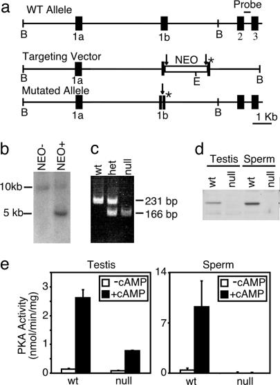
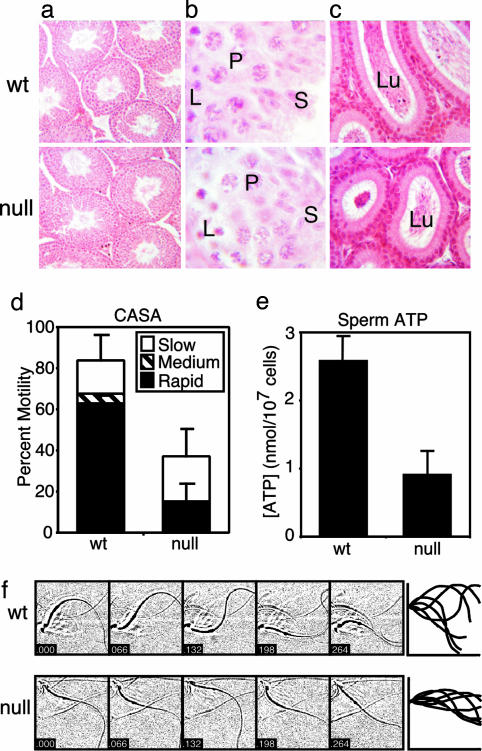
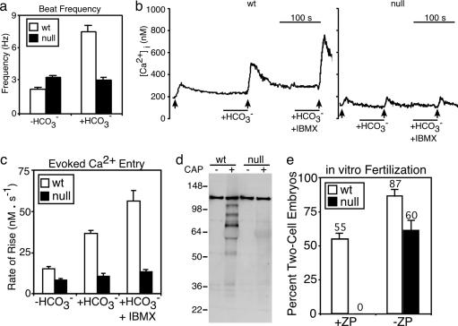
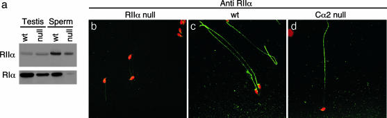
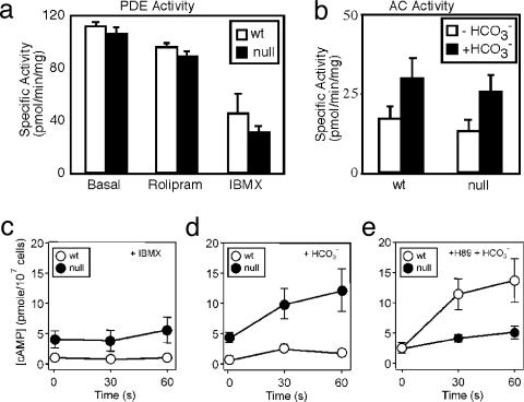
An unusual cAMP signaling system mediates many of the events that prepare spermatozoa to meet the egg. Its components include the atypical, bicarbonate-stimulated, sperm adenylyl cyclase and a cAMP-dependent protein kinase (PKA) with the unique catalytic subunit termed Calpha(2) or C(s). We generated mice that lack Calpha(2) to determine its importance in the events downstream of cAMP production. Male Calpha(2) null mice produce normal numbers of sperm that swim spontaneously in vitro. Thus, Calpha(2) has no required role in formation of a functional flagellum or the initiation of motility. In contrast, we find that Calpha(2) is required for bicarbonate to speed the flagellar beat and facilitate Ca(2+) entry channels. In addition, Calpha(2) is needed for the protein tyrosine phosphorylation that occurs late in the sequence of sperm maturation and for a negative feedback control of cAMP production, revealed here. Consistent with these specific defects in several important sperm functions, Calpha(2) null males are infertile despite normal mating behavior. These results define several crucial roles of PKA in sperm cell biology, bringing together both known and unique PKA-mediated events that are necessary for male fertility.


Figures

Fig. 1.
Fig. 1.
Targeted disruption of Cα2. (a) Schematic representation of the Cα gene, targeting vector, and mutated Cα2 allele. A loxP (→)-flanked NEO cassette and a mutation in the initiation codon (*) were inserted into exon 1b. The probe for Southern blot analysis is indicated between exons 2 and 3. Restriction enzyme sites shown are BamH1 (B) and EcoR1 (E). (b) Southern blot analysis of EcoR1-digested genomic DNA from the offspring of germ-line chimeric mice without (NEO–) or with (NEO+) the targeted NEO-containing allele. (c) PCR genotyping of genomic DNA after Cre-recombinase-mediated removal of the NEO cassette. The WT allele yielded a 231-bp product, and the mutated allele yielded a 166-bp product. (d) Western blot analysis of testis extracts from WT and homozygous null mice using a polyclonal antibody raised against the first 10 aa of Cα2.(e) In vitro kinase assay of testis and sperm extracts measuring basal (–cAMP) or total (+cAMP) PKA activity.
Fig. 2.
Fig. 2.
Testis and sperm analysis for Cα2 null mice. Histological comparison of stained WT and null tissue sections: (a) testis sections at low magnification (×200); (b) higher magnification (×1,000) of a stage IX-X tubule with labeled leptotene spermatocytes (L), pachytene spermatocytes (P), and elongating spermatids (S); and (c) caudae epidymides showing the lumen (Lu) of the tubules (×200). (d) Computer-assisted semen analysis (CASA) of motility of WT and null epididymal sperm examined after 30 min in capacitating conditions. Distributions of progressive motility were categorized as slow, medium, or fast. n = 3 mice per genotype, >200 cells per mouse. (e) ATP content of WT and null sperm. n = 3 mice per genotype. (f Left) Stop-motion images (48 × 48 μm) of WT and null sperm in HS medium (without formula image) collected at 66-ms intervals. (Right) Flagellar traces for one complete beat cycle from the cells shown to the left, after rotation to a horizontal axis and alignment to a common origin.
Fig. 3.
Fig. 3.
Absence of bicarbonate-stimulated and capacitation-dependent events in Cα2 null sperm. (a) Flagellar beat frequency of WT (n = 8) and null (n = 11) sperm. (b) Representative traces of indo-1 ratio-photometric monitoring of intracellular free [Ca2+] for WT and null sperm perfused with HS medium alone, then supplemented with 15 mM NaHCO3, then with 15 mM NaHCO3 and 200 μM IBMX. The ↑ indicate 10 s of depolarizing stimulus with K8.6 medium. (c) Conditioning with formula image facilitates the rate of Ca2+ entry determined from the averaged linear rising phase of responses as in b (WT n = 11, null n = 14). (d) Western blot analysis of phosphotyrosine content from WT and null sperm examined before (CAP–) or after (CAP+) a 90-min exposure to capacitating conditions. (e) In vitro fertilization of WT oocytes [+ or – zona pellucida (ZP)] using WT or null sperm. The percentage of two-cell embryos was determined 24 h after addition of sperm. Data represent the means of three independent experiments involving 75–145 oocytes, n = 3–5 male mice for each genotype.
Fig. 4.
Fig. 4.
Expression and localization of RI subunits in Cα2 null sperm. (a) Western blot analysis of extracts of WT and Cα2 null testis and sperm, probed for RIIα and RIα protein. (bd) Immunocytochemical localization of RIIα protein (green) in fixed cauda epididymal sperm from RIIα null mice (negative control) (b), WT mice (c), and Cα2 null mice (d). Sperm heads were stained with propidium iodide (red).
Fig. 5.
Fig. 5.
PKA-mediated feedback control of sAC. Total basal PDE assayed in extracts of WT and Cα2 null sperm. (a) Sensitivity of PDE activity to rolipram (20 μM) and IBMX (200 μM). (b) AC activity in sperm extracts assayed in the presence and absence of 15 mM NaHCO3.(ce) cAMP content was monitored over a 1-min time course for WT (○) and Cα2 null (•) sperm in HS medium supplemented at t = 0 with 200 μM IBMX (c), 15 mM NaHCO3 (d), or 15 mM NaHCO3 with 30 μM H89 applied after a 10-min exposure to 30 μM H89 alone (e). n = 3–6 mice per genotype.

References

    1. Boatman, D. E. & Robbins, R. S. (1991) Biol. Reprod. 44, 806–813. - PubMed
    1. Okamura, N., Tajima, Y., Soejima, A., Masuda, H. & Sugita, Y. (1985) J. Biol. Chem. 260, 9699–9705. - PubMed
    1. Gadella, B. M. & Harrison, R. A. (2000) Development (Cambridge, U.K.) 127, 2407–2420. - PubMed
    1. Carlson, A. E., Westenbroek, R. E., Quill, T., Ren, D., Clapham, D. E., Hille, B., Garbers, D. L. & Babcock, D. F. (2003) Proc. Natl. Acad. Sci. USA 100, 14864–14868. - PMC - PubMed
    1. Wennemuth, G., Carlson, A. E., Harper, A. J. & Babcock, D. F. (2003) Development (Cambridge, U.K.) 130, 1317–1326. - PubMed

Publication types

MeSH terms