

Understanding the mechanisms of drug-associated interstitial lung disease
- PMID: 15340376
- PMCID: PMC2750813
- DOI: 10.1038/sj.bjc.6602065
Understanding the mechanisms of drug-associated interstitial lung disease
Abstract
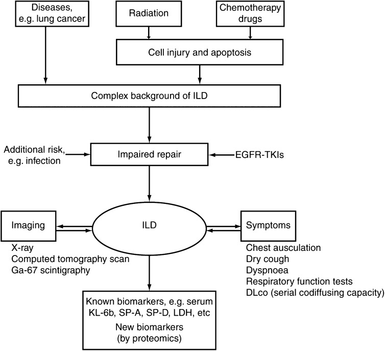
Drugs have been implicated in lung injury as a result of direct pharmacological action, persistence or metabolism in the tissue, or via the production of a reactive metabolite or metabolites. The result of this apparent drug-associated injury ranges from cellular dysfunction through to cell death (apoptosis) and alteration of repair mechanisms that are essential in replacing critical tissue elements and function. There is limited knowledge on how timing of drug administration or drug interactions may interfere with the repair mechanisms or modulate the expression of pulmonary toxicity. Chemotherapeutic drugs and novel agents, such as those targeting the epidermal growth factor receptor (EGFR), appear to affect both normal and neoplastic cells. However, unlike chemotherapy, where the actions are systemic and directly as a result of biotransformation or cell injury, it has been postulated that effects of EGFR-targeting agents are more likely to be focused on epithelia via a pharmacological effect. Furthermore, risk factors for the development of adverse pulmonary reactions, as well as biological markers indicating incipient toxicity, need to be prospectively identified. Proteomics, through the identification of >/=1000 proteins or peptides in blood samples, will hopefully identify candidates for this role.
Figures

Similar articles
-
EGFR-TK1-Associated Interstitial Pneumonitis in Nivolumab-Treated Patients with NSCLC: When Is it Worth The Risk?Pathol Oncol Res. 2019 Oct;25(4):1665-1666. doi: 10.1007/s12253-019-00599-z. Epub 2019 Jan 19. Pathol Oncol Res. 2019. PMID: 30661223 No abstract available.
-
Interstitial lung disease associated with epidermal growth factor receptor tyrosine kinase inhibitor therapy in non-small-cell lung carcinoma.Clin Lung Cancer. 2006 Dec;8 Suppl 1:S31-5. doi: 10.3816/clc.2006.s.011. Clin Lung Cancer. 2006. PMID: 17239288 Review.
-
EGFR-TKI-Associated Interstitial Pneumonitis in Nivolumab-Treated Patients With Non-Small Cell Lung Cancer.JAMA Oncol. 2018 Aug 1;4(8):1112-1115. doi: 10.1001/jamaoncol.2017.4526. JAMA Oncol. 2018. PMID: 29327061 Free PMC article.
-
The role of pharmacoethnicity in the development of cytotoxic and molecular targeted drugs in oncology.Yonsei Med J. 2013 Jan 1;54(1):1-14. doi: 10.3349/ymj.2013.54.1.1. Yonsei Med J. 2013. PMID: 23225792 Free PMC article. Review.
-
Fatal interstitial lung disease associated with gemcitabine and erlotinib therapy for lung cancer.Med Oncol. 2012 Mar;29(1):212-4. doi: 10.1007/s12032-010-9790-y. Epub 2011 Jan 25. Med Oncol. 2012. PMID: 21264548
Cited by
-
Drug-induced interstitial lung disease: mechanisms and best diagnostic approaches.Respir Res. 2012 May 31;13(1):39. doi: 10.1186/1465-9921-13-39. Respir Res. 2012. PMID: 22651223 Free PMC article. Review.
-
Molecular mechanisms of hexavalent chromium-induced apoptosis in human bronchoalveolar cells.Am J Respir Cell Mol Biol. 2005 Dec;33(6):589-600. doi: 10.1165/rcmb.2005-0213OC. Epub 2005 Sep 15. Am J Respir Cell Mol Biol. 2005. PMID: 16166740 Free PMC article.
-
Severe eosinophilic pneumonia presenting during gemcitabine adjuvant chemotherapy.World J Surg Oncol. 2013 Jul 24;11:167. doi: 10.1186/1477-7819-11-167. World J Surg Oncol. 2013. PMID: 23883337 Free PMC article.
-
Epidermal growth factor receptor inhibitors in non-small cell lung cancer.Drugs. 2007;67(8):1125-38. doi: 10.2165/00003495-200767080-00003. Drugs. 2007. PMID: 17521215 Review.
-
Effects of neoadjuvant chemotherapy on respiratory function in patients with breast cancer.Chin J Cancer Res. 2020 Feb;32(1):36-42. doi: 10.21147/j.issn.1000-9604.2020.01.05. Chin J Cancer Res. 2020. PMID: 32194303 Free PMC article.
References
-
- Abid SH, Malhotra V, Perry MC (2001) Radiation-induced and chemotherapy-induced pulmonary injury. Curr Opin Oncol 13: 242–248 - PubMed
-
- Ackerman AD, Fackler JC, Tuck-Muller CM, Tarpey MM, Freeman BA, Rogers MC (1988) Partial monosomy 21, diminished activity of superoxide dismutase, and pulmonary oxygen toxicity. N Engl J Med 318: 1666–1669 - PubMed
-
- Aida S, Tamai S, Sekiguchi S, Shimizu N (1994) Distribution of epidermal growth factor and epidermal growth factor receptor in human lung: immunohistochemical and immunoelectron-microscopic studies. Respiration 61: 161–166 - PubMed
-
- Anttila S, Hietanen E, Vainio H, Camus AM, Gelboin HV, Park SS, Heikkila L, Karjalainen A, Bartsch H (1991) Smoking and peripheral type of cancer are related to high levels of pulmonary cytochrome P450IA in lung cancer patients. Int J Cancer 47: 681–685 - PubMed
-
- Aviram G, Yu E, Tai P, Lefcoe MS (2001) Computed tomography to assess pulmonary injury associated with concurrent chemo-radiotherapy for inoperable non-small cell lung cancer. Can Assoc Radiol J 52: 385–391 - PubMed
Publication types
MeSH terms
Substances
LinkOut - more resources
Full Text Sources
Medical
Research Materials
Miscellaneous
