

Cellular antitumor immune response to a branched lysine multiple antigenic peptide containing epitopes of a common tumor-specific antigen in a rat glioma model
- PMID: 15340764
- PMCID: PMC11032903
- DOI: 10.1007/s00262-004-0576-y
Cellular antitumor immune response to a branched lysine multiple antigenic peptide containing epitopes of a common tumor-specific antigen in a rat glioma model
Abstract
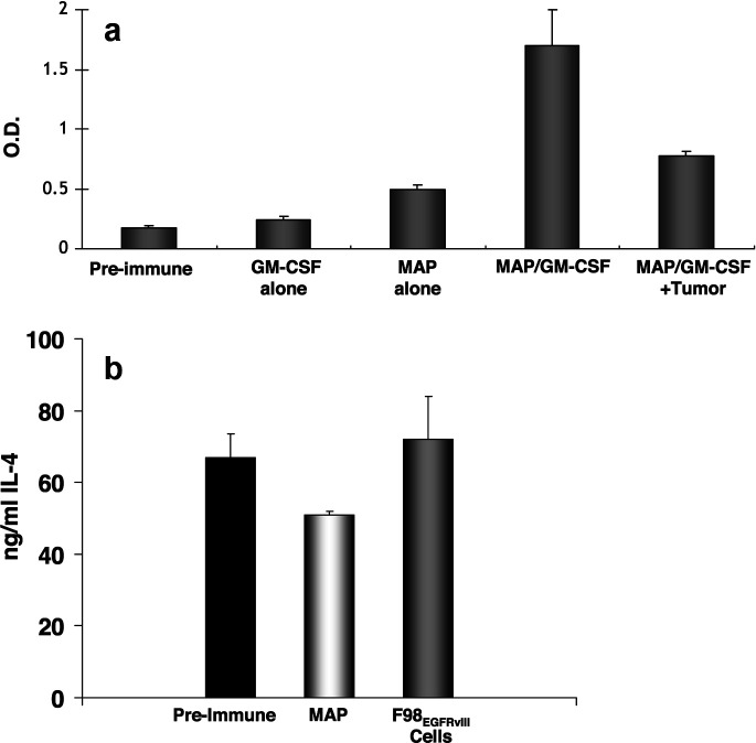
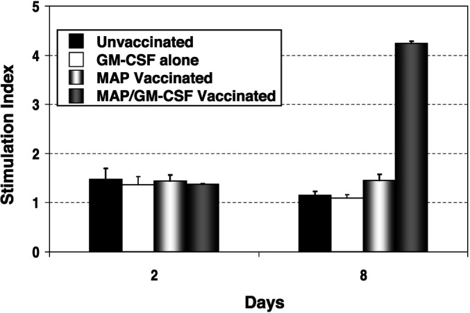
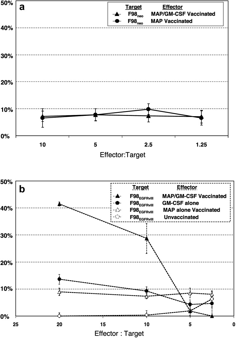
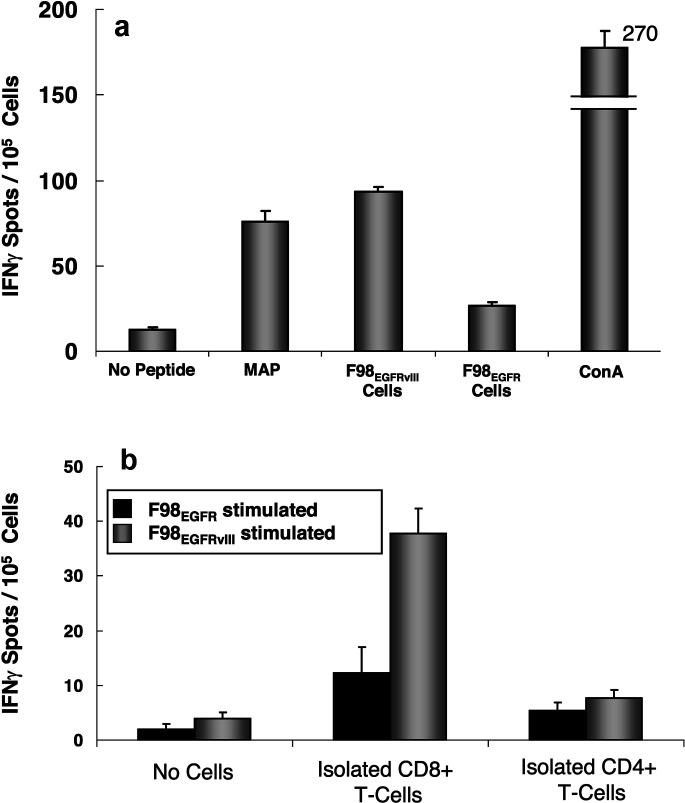
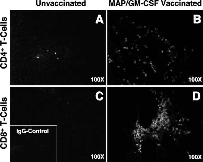
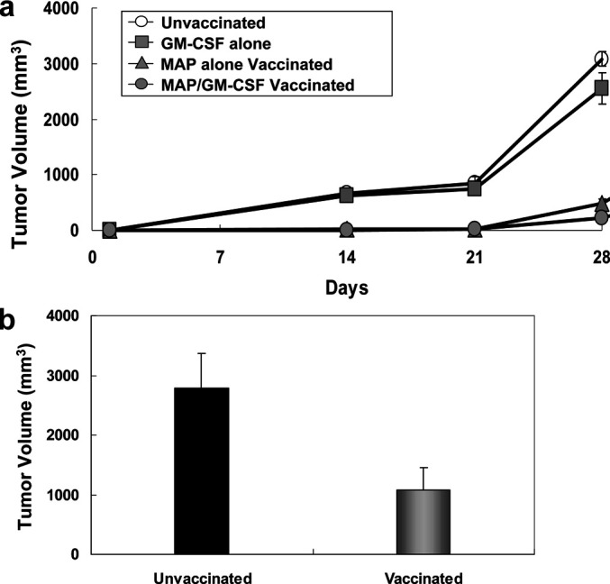
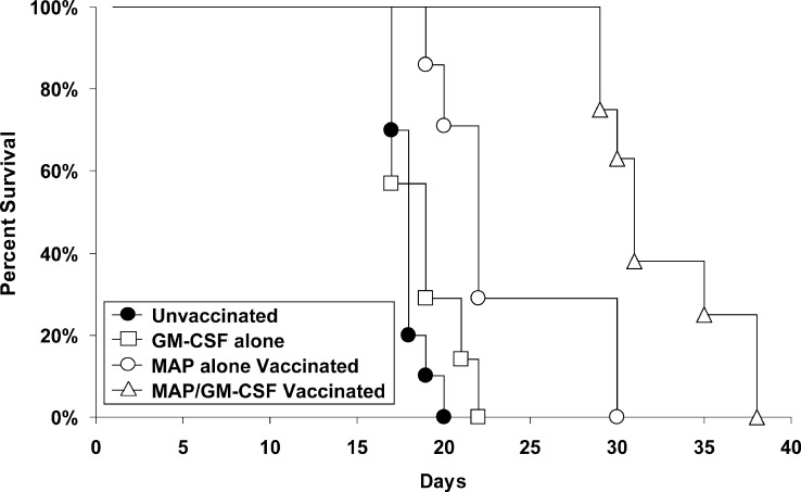
Human malignant gliomas contain epidermal growth factor receptor (EGFR) gene mutations that encode tumor-associated antigens (TAAs) that can be targeted using immunological techniques. One EGFR mutant gene (EGFRvIII) encodes a protein with an epitope that is not found in normal tissues. A number of studies have focused on this unique epitope as a potential target for tumor vaccines. In the present study, we examined the cellular immune effects of a peptide containing multiple copies of the unique EGFRvIII epitope linked together by way of a lysine bridge. Fischer rats were vaccinated with an EGFRvIII multiple antigenic peptide (MAP). While vaccination produced a humoral immune response, anti-MAP antibody production was not accompanied by expression of the Th2 response cytokine IL-4. In MAP/GM-CSF vaccinated animals, a cellular immune response was detected in association with the appearance of CD4+ and CD8+ T cells at the tumor site. Splenocytes and CD8+ T cells from vaccinated rats produced the Th1 cytokine IFN-gamma in vitro in response to stimulation by rat glioma cells expressing EGFRvIII, but not by those expressing wild-type EGFR. MAP vaccine also induced a specific lytic antitumor CTL immune response against F98 glioma cells expressing EGFRvIII, but not against F98 cells expressing either wild-type EGFR or no receptor. The in vivo growth of F98(EGFRvIII) cells was attenuated in vaccinated rats; whereas, growth of F98(EGFR) cells was not. The median survival of vaccinated rats was increased 72% over that of unvaccinated controls challenged with intracerebral F98(EGFRvIII) tumor implants. Therefore, MAP vaccination produced a predominantly cellular antitumor immune response directed against F98 gliomas expressing the EGFRvIII target antigen. The potent immunosuppressive effects of F98 glioma cells mimic the human disease and make this particular tumor model useful for studying immunotherapeutic approaches to malignant gliomas.
Figures


Similar articles
-
Generation of anti-idiotypic reagents in the EGFRvIII tumor-associated antigen system.Cancer Immunol Immunother. 2002 Feb;50(12):639-52. doi: 10.1007/s00262-001-0243-5. Epub 2001 Dec 8. Cancer Immunol Immunother. 2002. PMID: 11862416 Free PMC article.
-
Sequential immunotherapy by vaccination with GM-CSF-expressing glioma cells and CTLA-4 blockade effectively treats established murine intracranial tumors.J Immunother. 2012 Jun;35(5):385-9. doi: 10.1097/CJI.0b013e3182562d59. J Immunother. 2012. PMID: 22576343 Free PMC article.
-
Glioma-specific cytotoxic T cells can be effectively induced by subcutaneous vaccination of irradiated wild-type tumor cells without artificial cytokine production.Int J Oncol. 2003 Aug;23(2):483-8. Int J Oncol. 2003. PMID: 12851699
-
Tumor-specific immunotherapy targeting the EGFRvIII mutation in patients with malignant glioma.Semin Immunol. 2008 Oct;20(5):267-75. doi: 10.1016/j.smim.2008.04.001. Epub 2008 Jun 9. Semin Immunol. 2008. PMID: 18539480 Free PMC article. Review.
-
The evolution of the EGFRvIII (rindopepimut) immunotherapy for glioblastoma multiforme patients.Hum Vaccin Immunother. 2014;10(11):3322-31. doi: 10.4161/21645515.2014.983002. Hum Vaccin Immunother. 2014. PMID: 25625931 Free PMC article. Review.
Cited by
-
Targeted therapies for malignant glioma: progress and potential.BioDrugs. 2009;23(1):25-35. doi: 10.2165/00063030-200923010-00003. BioDrugs. 2009. PMID: 19344189 Free PMC article. Review.
-
Antitumor effects of a xenogeneic survivin bone marrow derived dendritic cell vaccine against murine GL261 gliomas.Cancer Immunol Immunother. 2006 Dec;55(12):1491-503. doi: 10.1007/s00262-006-0138-6. Epub 2006 Feb 17. Cancer Immunol Immunother. 2006. PMID: 16485128 Free PMC article.
-
Development of Peptide-Based Vaccines for Cancer.J Oncol. 2022 Mar 15;2022:9749363. doi: 10.1155/2022/9749363. eCollection 2022. J Oncol. 2022. PMID: 35342400 Free PMC article. Review.
-
DNA prime and peptide boost immunization protocol encoding the Toxoplasma gondii GRA4 induces strong protective immunity in BALB/c mice.BMC Infect Dis. 2013 Oct 23;13:494. doi: 10.1186/1471-2334-13-494. BMC Infect Dis. 2013. PMID: 24148219 Free PMC article.
-
Glioma diagnostics and biomarkers: an ongoing challenge in the field of medicine and science.Expert Rev Mol Diagn. 2014 May;14(4):439-52. doi: 10.1586/14737159.2014.905202. Expert Rev Mol Diagn. 2014. PMID: 24746164 Free PMC article. Review.
References
-
- Wingo PA, Tong T, Bolden S. Cancer statistics. CA Cancer J Clin. 1995;45:8–30. - PubMed
-
- McKinley B, Michalek A, Plunkett RJ, Fenstermaker RA. The impact of age and sex on the incidence of glial tumors in New York State from 1976 to 1995. J Neurosurg. 2000;93:932–939. - PubMed
-
- Libermann TA, Nusbaum HR, Razon N, et al. Amplification, enhanced expression and possible rearrangement of EGF receptor gene in primary human brain tumours of glial origin. Nature. 1985;313:144–147. - PubMed
Publication types
MeSH terms
Substances
Grants and funding
LinkOut - more resources
Full Text Sources
Other Literature Sources
Research Materials
Miscellaneous
