

Titanium mesh cages (TMC) in spine surgery
- PMID: 15340827
- PMCID: PMC3476740
- DOI: 10.1007/s00586-004-0748-7
Titanium mesh cages (TMC) in spine surgery
Abstract
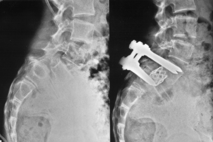
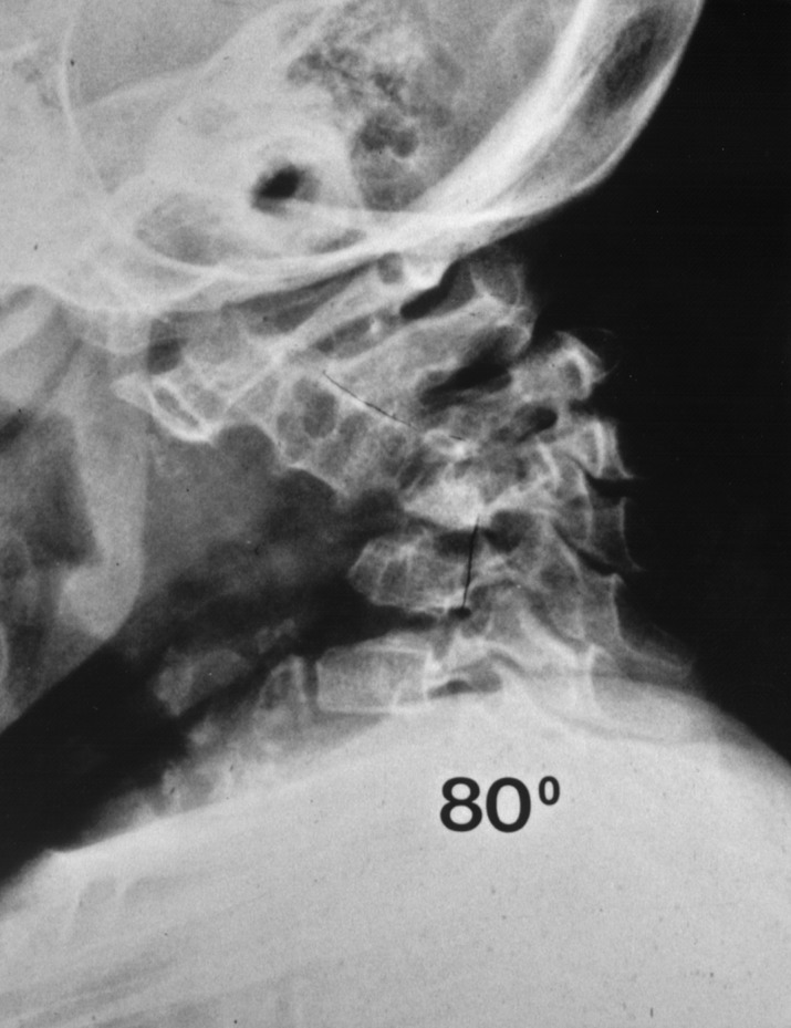
The introduction of the titanium mesh cage (TMC) in spinal surgery has opened up a variety of applications that are realizable as a result of the versatility of the implant. Differing applications of TMCs in the whole spine are described in a series of 150 patients. Replacement and reinforcement of the anterior column represent the classic use of cylindrical TMCs. The TMC as a multisegmental concave support in kyphotic deformities and as a posterior interlaminar spacer or lamina replacement after wide laminectomy are additional applications. Implant subsidence, pseudarthrosis and implant loosening are the complications typically encountered with use of TMCs. The versatility of the implant permits its use in unusual surgical situations.
Figures


Similar articles
-
Effect of posterior subsidence on cervical alignment after anterior cervical corpectomy and reconstruction using titanium mesh cages in degenerative cervical disease.J Clin Neurosci. 2014 Oct;21(10):1779-85. doi: 10.1016/j.jocn.2014.02.016. Epub 2014 Jun 21. J Clin Neurosci. 2014. PMID: 24957629
-
Titanium mesh cages for cervical spine stabilization after corpectomy: a clinical and radiological study.J Neurosurg. 2003 Sep;99(2 Suppl):172-80. doi: 10.3171/spi.2003.99.2.0172. J Neurosurg. 2003. PMID: 12956460
-
Effectiveness of titanium mesh cylindrical cages in anterior column reconstruction after thoracic and lumbar vertebral body resection.Spine (Phila Pa 1976). 2003 May 1;28(9):902-8. doi: 10.1097/01.BRS.0000058712.88053.13. Spine (Phila Pa 1976). 2003. PMID: 12942006
-
Mesh cages for spinal deformity in adults.Clin Orthop Relat Res. 2002 Jan;(394):92-7. doi: 10.1097/00003086-200201000-00011. Clin Orthop Relat Res. 2002. PMID: 11795756 Review.
-
The use of titanium mesh cages in the cervical spine.Clin Orthop Relat Res. 2002 Jan;(394):47-54. doi: 10.1097/00003086-200201000-00006. Clin Orthop Relat Res. 2002. PMID: 11795751 Review.
Cited by
-
Customized Multilevel 3D Printing Implant for Reconstructing Spine Tumor: A Retrospective Case Series Study in a Single Center.Orthop Surg. 2022 Sep;14(9):2016-2022. doi: 10.1111/os.13357. Epub 2022 Jul 27. Orthop Surg. 2022. PMID: 35894154 Free PMC article.
-
Surgical Strategy to Protect the Exposed Spinal Cord From Extrinsic Compression in Severe Kyphosis: A Case Report.HSS J. 2022 Feb;18(1):166-170. doi: 10.1177/1556331621993063. Epub 2021 Feb 19. HSS J. 2022. PMID: 35087347 Free PMC article. No abstract available.
-
Biomechanical Effects of Different Prosthesis Types and Fixation Ranges in Multisegmental Total En Bloc Spondylectomy: A Finite Element Study.Orthop Surg. 2024 Oct;16(10):2488-2498. doi: 10.1111/os.14171. Epub 2024 Aug 5. Orthop Surg. 2024. PMID: 39101231 Free PMC article.
-
Mineralized collagen-modified PMMA cement enhances bone integration and reduces fibrous encapsulation in the treatment of lumbar degenerative disc disease.Regen Biomater. 2020 Mar;7(2):181-193. doi: 10.1093/rb/rbz044. Epub 2019 Dec 2. Regen Biomater. 2020. PMID: 32296537 Free PMC article.
-
Thermal Localization Improves the Interlayer Adhesion and Structural Integrity of 3D printed PEEK Lumbar Spinal Cages.Materialia (Oxf). 2020 May;10:100650. doi: 10.1016/j.mtla.2020.100650. Epub 2020 Mar 9. Materialia (Oxf). 2020. PMID: 32318685 Free PMC article.
References
Publication types
MeSH terms
Substances
LinkOut - more resources
Full Text Sources
Medical
