. 2004 Sep;12(9):1607-18.
doi: 10.1016/j.str.2004.06.019.

Conformational changes of the flavivirus E glycoprotein

Affiliations

Conformational changes of the flavivirus E glycoprotein

Ying Zhang et al. Structure. 2004 Sep.

Abstract

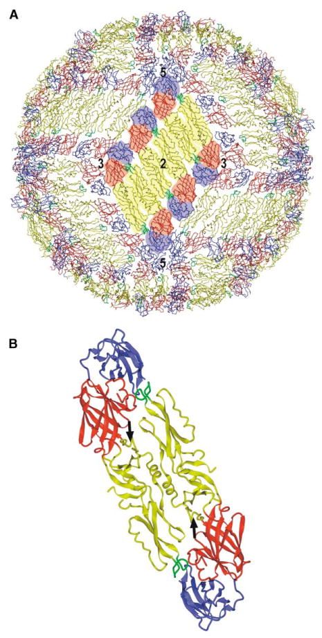
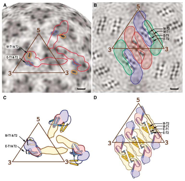
Dengue virus, a member of the Flaviviridae family, has a surface composed of 180 copies each of the envelope (E) glycoprotein and the membrane (M) protein. The crystal structure of an N-terminal fragment of E has been determined and compared with a previously described structure. The primary difference between these structures is a 10 degrees rotation about a hinge relating the fusion domain DII to domains DI and DIII. These two rigid body components were used for independent fitting of E into the cryo-electron microscopy maps of both immature and mature dengue viruses. The fitted E structures in these two particles showed a difference of 27 degrees between the two components. Comparison of the E structure in its postfusion state with that in the immature and mature virions shows a rotation approximately around the same hinge. Flexibility of E is apparently a functional requirement for assembly and infection of flaviviruses.


Figures

Figure 1
Figure 1. The Mature Dengue Virus Structure
(A) Packing of the E proteins in the mature virus, showing the herringbone pattern. One of the 30 rafts, each containing three parallel dimers, is highlighted. Domains I, II, and III are colored red, yellow, and blue, respectively, with the fusion peptide in green. Symmetry axes are labeled. (B) Ribbon diagram of the crystal dimer with the same color coding as in (A). Black arrows point to the kl β-hairpin in each monomer, where β-OG can bind.
Figure 2
Figure 2. Sequence Alignment of TBEV and DEN sE Based on Structural Comparisons
The numbering system is from DEN. The surface accessibility of the DEN sE(P) dimer, averaged over an 11 residue window, is plotted in violet. The temperature factor of DEN sE(P) is shown in dark blue. The plot of residue conservation between TBEV and DEN, measured in terms of point-accepted mutations (PAM) (Dayhoff, 1972) and averaged over an 11 residue window, is shown in light blue. The plot of Cα differences (brown) between TBEV and DEN sE is also shown. Black bars indicate regions with low sequence conservation. Residues of domains I, II, and III are in red, yellow, and blue, respectively. The four hinge peptides linking domains DI and DII are, therefore, at the red-yellow boundaries. Note the presence of conserved glycines in the hinge region. Secondary structural elements are shown below the sequences.
Figure 3
Figure 3. Conformational Changes of the sE Structure
(A and B) Stereo diagram showing superposition of TBEV (magenta), DEN sE(P) (yellow), and DEN sE(H) without β-OG (red) for (A) DI + DIII and (B) DII, viewed along the direction of the dimer 2-fold axis. (C) Stereoview of superimposed E ectodomains showing their different hinge angles along the direction of the hinge axis, perpendicular to the view in (A) and (B). The alignment is based on DI, and the diagrammatic position of DIII corresponds to the prefusion structure.
Figure 4
Figure 4. The E Protein Structure in the Mature Virus
(A) Stereoview of the E protein fitted to the cryo-EM density of the mature DEN around the 5-fold axis. Domains DI, DII, and DIII are colored as in Figure 1. The AxA, CD, and FG surface loops are shown as Cα traces. (B) Stereo diagram showing sE fitted in the cryo-EM electron density map of the mature DEN virus. Molecules 1, 2, and 3 (see Table 3) are in red, green, and blue, respectively. Symmetry axes are labeled. (C) Side view of the E dimer as determined by the independent fitting of DI + DIII and of DII into the density of the mature virus (red) superimposed onto the sE(P) dimer in the crystal (yellow). Note that the colors in (A) compared to (B) and (C) in both Figures 4 and 5 represent different structural components.
Figure 5
Figure 5. The E Protein Structure in the Immature Virus
(A) Stereo diagram showing sE fitted into the cryo-EM electron density map of immature DEN. Molecules 1, 2, and 3 (see Table 3) are in red, green, and blue, respectively. Symmetry axes are labeled. (B) Stereoview of the Cα trace of domains DI (red), DII (yellow), DIII (blue), and the stem-anchor region (violet) of E in the map of the immature virus. The electron density that corresponds to the membrane is colored green. (C) The alignment of the E proteins in the mature and immature viruses. The superposition is based on DIII of the sE fragment. The ectodomain is shown as in (B). The stem-anchor regions in mature and immature viruses are colored cyan and violet, respectively. The C terminus of the E ectodomain is marked with “C.” Note the similar positions of helix H1.
Figure 6
Figure 6. The Structure of the Stem and Anchor Regions
Position of the stem and anchor regions of the E and M proteins in the immature (A and C) and mature (B and D) particles. Radial projections of the three-dimensional cryo-EM densities at radius of 180 Å is shown for the (A) immature and (B) mature dengue virus particles, corresponding roughly to the middle of the lipid bilayer. The outlines of three E monomers, representing one asymmetric unit, are shown in red in (A). The three E monomers per asymmetric unit, arranged as a raft of dimers, are colored red, blue, and green in (B). Darker shading represents higher density regions resulting from the transmembrane protein helices E-T1 and E-T2 of the E glycoprotein and M-T1 and M-T2 for the membrane protein. Structural interpretations are shown in (C) and (D), respectively, where the color code for domains I, II, and III of the E protein is the same as in Figure 1. The scale bar is 20 Å long.

Similar articles

Cited by

References

    1. Allison SL, Schalich J, Stiasny K, Mandl CW, Kunz C, Heinz FX. Oligomeric rearrangement of tick-borne encephalitis virus envelope proteins induced by an acidic pH. J Virol. 1995;69:695–700. - PMC - PubMed
    1. Allison SL, Schalich J, Stiasny K, Mandl CW, Heinz FX. Mutational evidence for an internal fusion peptide in flavivirus envelope protein E. J Virol. 2001;75:4268–4275. - PMC - PubMed
    1. Bhardwaj S, Holbrook M, Shope RE, Barrett ADT, Watowich SJ. Biophysical characterization and vector-specific antagonist activity of domain III of the tick-borne flavivirus envelope protein. J Virol. 2001;75:4002–4007. - PMC - PubMed
    1. Bressanelli S, Stiasny K, Allison SL, Stura EA, Duquerroy S, Lescar J, Heinz FX, Rey FA. Structure of a flavivirus envelope glycoprotein in its low-pH-induced membrane fusion conformation. EMBO J. 2004;23:728–738. - PMC - PubMed
    1. Brünger AT, Adams PD, Clore GM, DeLano WL, Gros P, Grosse-Kunstleve RW, Jiang JS, Kuszewski J, Nilges M, Pannu NS, et al. Crystallography and NMR system: a new software suite for macromolecular structure determination. Acta Crystallogr D Biol Crystallogr. 1998;54:905–921. - PubMed

Publication types

Substances

LinkOut - more resources