Comparative Study
. 2004 Sep 1;18(17):2134-46.
doi: 10.1101/gad.1214104.

Attenuation of estrogen receptor alpha-mediated transcription through estrogen-stimulated recruitment of a negative elongation factor

Affiliations
Comparative Study

Attenuation of estrogen receptor alpha-mediated transcription through estrogen-stimulated recruitment of a negative elongation factor

Sarah E Aiyar et al. Genes Dev. .

Abstract

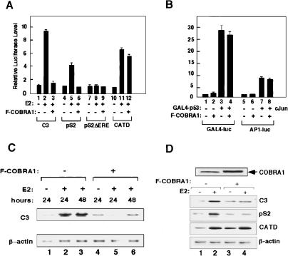
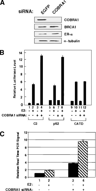
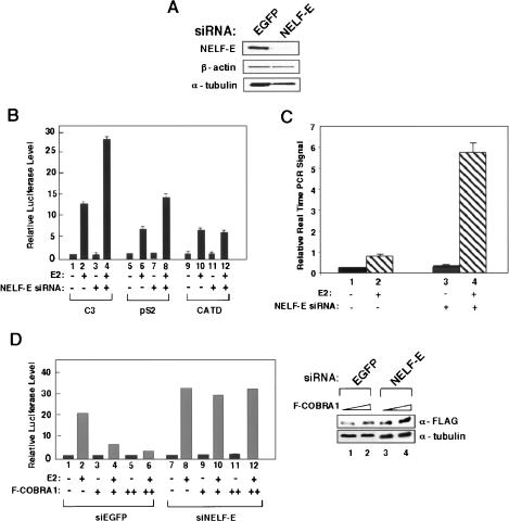
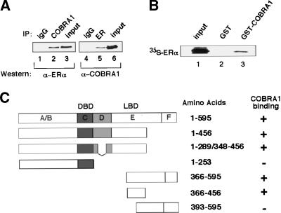
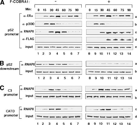
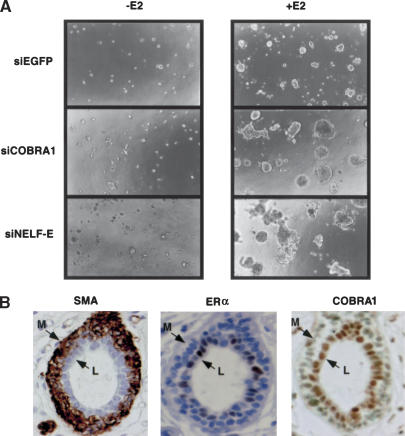
Estrogen receptor alpha (ERalpha) signaling is paramount for normal mammary gland development and function and the repression of breast cancer. ERalpha function in gene regulation is mediated by a number of coactivators and corepressors, most of which are known to modify chromatin structure and/or influence the assembly of the regulatory complexes at the level of transcription initiation. Here we describe a novel mechanism of attenuating the ERalpha activity. We show that cofactor of BRCA1 (COBRA1), an integral subunit of the human negative elongation factor (NELF), directly binds to ERalpha and represses ERalpha-mediated transcription. Reduction of the endogenous NELF proteins in breast cancer cells using small interfering RNA results in elevated ERalpha-mediated transcription and enhanced cell proliferation. Chromatin immunoprecipitation reveals that recruitment of COBRA1 and the other NELF subunits to endogenous ERalpha-responsive promoters is greatly stimulated upon estrogen treatment. Interestingly, COBRA1 does not affect the estrogen-dependent assembly of transcription regulatory complexes at the ERalpha-regulated promoters. Rather, it causes RNA polymerase II (RNAPII) to pause at the promoter-proximal region, which is consistent with its in vitro biochemical activity. Therefore, our in vivo work defines the first corepressor of nuclear receptors that modulates ERalpha-dependent gene expression by stalling RNAPII. We suggest that this new level of regulation may be important to control the duration and magnitude of a rapid and reversible hormonal response.


Figures

Figure 1.
Figure 1.
Ectopically expressed COBRA1 represses ligand-dependent transcriptional activation by ERα. (A) COBRA1 represses transcription from the luciferase-linked C3 and pS2 promoters, but not the CATD promoter. T47D cells were transfected with 0.5 μg COBRA1 and 0.5 μg of various luciferase reporter plasmids in the presence or absence of 10 nM E2. (B) COBRA1 does not affect transcription from ERα-unresponsive promoters. GAL4-p53 and c-Jun were cotransfected with a GAL4- or AP1-responsive luciferase reporter construct, respectively, in the presence or absence of Flag-COBRA1. (C) Northern hybridization indicates the impact of ectopic COBRA1 on the transcription of the endogenous C3 gene in T47D cells. Stably transfected T47D clones harboring either the pcDNA3 empty vector or Flag-COBRA1-expressing plasmid were treated with either ethanol or E2 for the indicated periods of time and harvested for RNA extraction. (D) RT–PCR analysis of the COBRA1 effect on the transcription of various ER-responsive genes. Pools of T47D cells stably transfected with either empty or COBRA1-expressing retroviral vectors were treated with ethanol or E2 (10 nM) for 24 h. RT–PCR was conducted on the total RNA to assess the transcription level of three ERα-responsive genes. In both C and D, β-Actin was used as an internal control. Also shown on the top of the panel is an anti-COBRA1 immunoblot indicating the total level of COBRA1 in the control and Flag-COBRA1-expressing cells.
Figure 2.
Figure 2.
Reduction of endogenous COBRA1 in T47D cells enhances gene activation at a subset of ERα-responsive promoters. (A) Immunoblots with various antibodies indicating the specific knockdown effect of the COBRA1 siRNA on the endogenous COBRA1 protein level. Quantitative immunoblotting indicates ∼90% reduction of the COBRA1 protein in the knockdown cells. (B) Luciferase reporter assays in the control (EGFP siRNA) and COBRA1 siRNA-expressing cells, using three ERα-responsive promoters. (C) Real-time PCR determining the pS2 transcript levels in the control and COBRA1 knockdown cells. Results are averages of triplicates that are normalized against the transcript levels of β-actin.
Figure 3.
Figure 3.
COBRA1-mediated transcriptional repression requires the other NELF subunits. (A) Immunoblots of the endogenous NELF-E, β-actin, and α-tubulin in the control and NELF-E knockdown cells. (B) Luciferase reporter assay using three ERα-responsive promoters in the control and NELF-E knockdown cells. (C) Real-time PCR determining the pS2 transcript levels in the control and NELF-E knockdown cells. Results are normalized against the transcript levels of β-actin. (D) The effect of ectopic COBRA1 on the C3-luciferase activity in the control (lanes 16) and NELF-E knockdown cells (lanes 712). Also shown on the right is an anti-Flag immunoblot, indicating the levels of ectopic COBRA1 in the + and ++ samples.
Figure 4.
Figure 4.
COBRA1 interacts with ERα in vivo and in vitro. (A) Interaction between the endogenous COBRA1 and ERα. Nuclear lysates of breast cancer cells (MCF7) were immunoprecipitated with an anti-ERα or anti-COBRA1 antibody. The immunoprecipitates were subsequently probed with anti-COBRA1 or anti-ERα antibody. (Lanes 1,4) Purified rabbit or mouse IgG was used as a negative control. (B) GST pull-down assay for detecting the in vitro interaction between GST-COBRA1 and in vitro translated ERα. (C) Diagram indicating the affinity of various ERα constructs for COBRA1. Results were obtained from both co-IP and GST pulldown assays.
Figure 5.
Figure 5.
Loading of endogenous NELF subunits at ERα-responsive promoters in a ligand-stimulated manner. (A) Ligand-stimulated recruitment of COBRA1, NELF-C/D, and NELF-E. T47D cells were treated with either ethanol or 10 nM E2 and subsequently cross-linked with formaldehyde. The cell lysates were used for the chromatin immunoprecipitation (ChIP) assay with anti-COBRA1 (rows ae), anti-NELF-E (rows f,g), or anti-NELF-C/D (rows h,i) antibody. DNA fragments corresponding to various promoter regions (indicated on right) were amplified by PCR from input (lanes 14) and ChIP samples (lanes 58). (B) Simultaneous recruitment of COBRA1, NELF-E, and NELF-C/D to the C3 and pS2 promoters. Cross-linked T47D cell lysates were precipitated with the indicated antibody. The immunoprecipitates were re-ChIPed with either IgG or the antibody against one of the other NELF subunits. (C) Time course study of the in vivo association of endogenous ERα or COBRA1 with the pS2 promoter region. T47D cells were harvested at various periods of time after the ligand treatment. ChIP analysis was conducted by using anti-ERα and COBRA1 antibodies. Also shown is a diagram of the pS2 gene that indicates the relative positions of the primers used in the ChIP assay in this study.
Figure 6.
Figure 6.
Ectopic expression of COBRA1 results in stalled RNAPII at the promoter-proximal region of the ERα-responsive promoters. (A) Occupancy by ERα, p300, RNAPII, and Flag-COBRA1 at the pS2 promoter at 15-min time intervals after the addition of E2 in either the empty vector (lanes 17) or Flag-COBRA1 (lanes 814) retroviral stable cell lines. (B) Time course of RNAP II occupancy ∼3 kb downstream of the pS2 promoter in the control and Flag-COBRA1-expressing cell lines. (C) Time course of RNAP II occupancy at the C3 and CATD promoters in the control and Flag-COBRA1-expressing cell lines.
Figure 7.
Figure 7.
NELF in breast cancer cell lines and normal breast tissues. (A) Control, COBRA1, and NELF-E siRNA knockdown cells were grown in the Matrigel-containing three-dimensional tissue culture system in the presence or absence of E2 (10 nM). After 3 wk of growth incubation, the images were observed under phase microscopy with a 10× objective. (B) COBRA is preferentially expressed in the luminal epithelial cells of the mammary gland. Immunostaining of normal mammary ducts for COBRA1 (right) and ERα (center). The exclusive staining for smooth muscle actin (SMA) in myoepithelial cells is shown as a control (left). The brown color indicates the presence of protein. The locations of luminal epithelial (L) and myoepithelial cells (M) are also indicated.

References

    1. Ainbinder E., Amir-Zilberstein, L., Yamaguchi, Y., Handa, H., and Dikstein, R. 2004. Elongation inhibition by DRB sensitivity-inducing factor is regulated by the A20 promoter via a novel negative element and NF-κB. Mol. Biol. Cell. 24: 2444-2454. - PMC - PubMed
    1. Ali S. and Coombes, R.C. 2002. Endocrine-responsive breast cancer and strategies for combating resistance. Nat. Rev. Cancer 2: 101-112. - PubMed
    1. Andrulis E.D., Guzman, E., Doring, P., Werner, J., and Lis, J.T. 2000. High-resolution localization of Drosophila Spt5 and Spt6 at heat shock genes in vivo: Roles in promoter proximal pausing and transcription elongation. Genes & Dev. 14: 2635-2649. - PMC - PubMed
    1. Augereau P., Miralles, F., Cavailles, V., Gaudelet, C., Parker, M., and Rochefort, H. 1994. Characterization of the proximal estrogen-responsive element of human cathepsin D gene. Mol. Endocrinol. 8: 693-703. - PubMed
    1. Baniahmad C., Nawaz, Z., Baniahmad, A., Gleeson, M.A., Tsai, M.J., and O'Malley, B.W. 1995. Enhancement of human estrogen receptor activity by SPT6: A potential coactivator. Mol. Endocrinol. 9: 34-43. - PubMed

Publication types

MeSH terms

LinkOut - more resources