Comparative Study
. 2004 Sep 1;18(17):2147-60.
doi: 10.1101/gad.302904.

Association of the transcriptional corepressor TIF1beta with heterochromatin protein 1 (HP1): an essential role for progression through differentiation

Affiliations
Comparative Study

Association of the transcriptional corepressor TIF1beta with heterochromatin protein 1 (HP1): an essential role for progression through differentiation

Florence Cammas et al. Genes Dev. .

Abstract

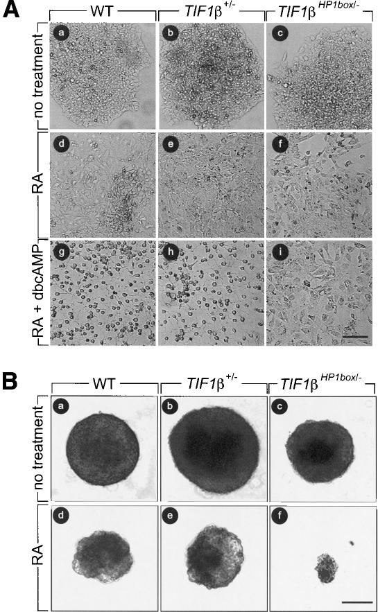
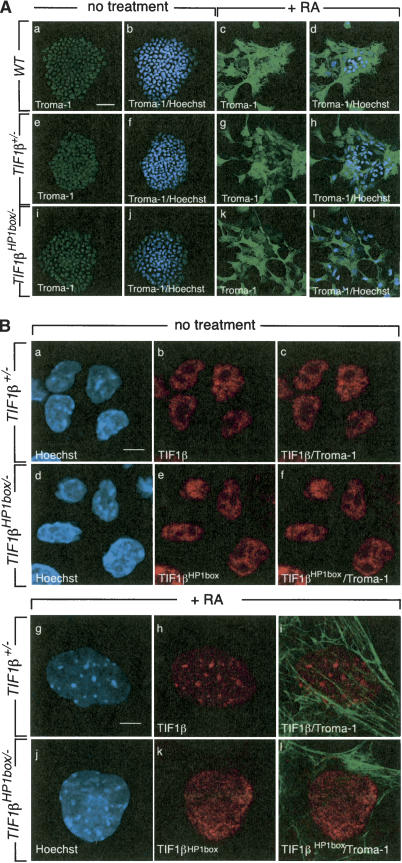
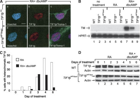
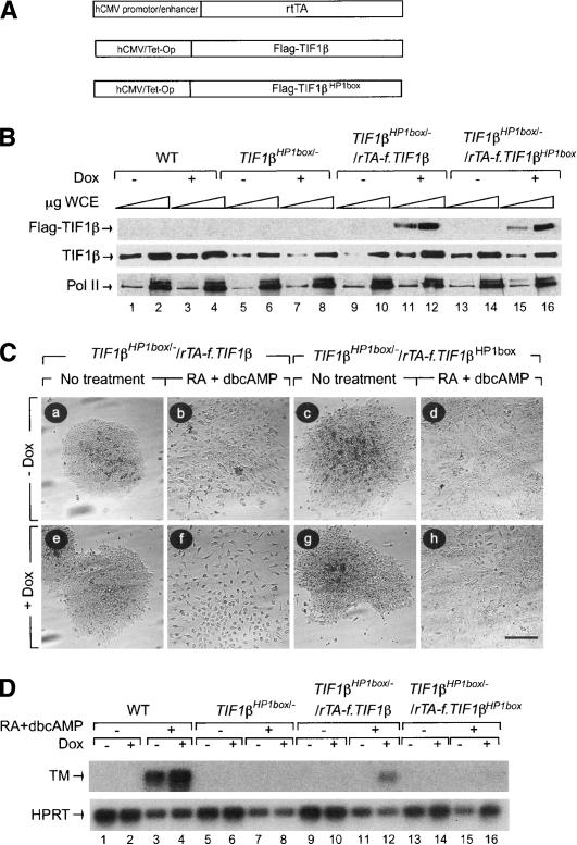
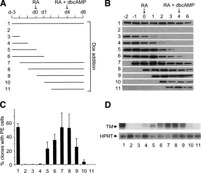
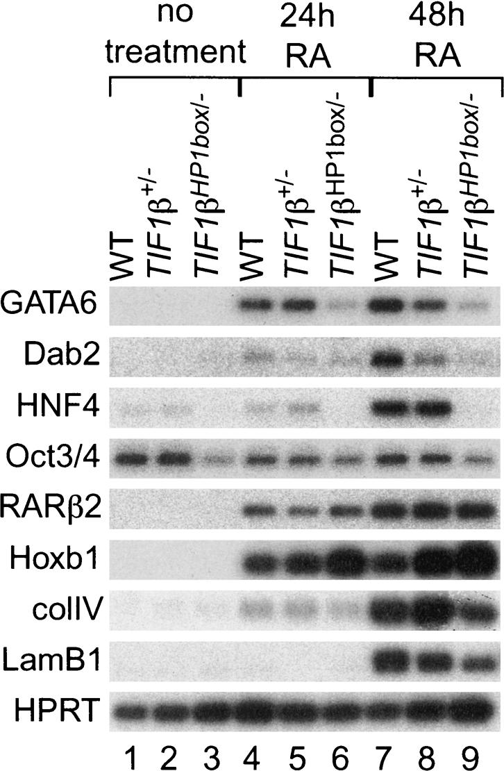
The transcriptional intermediary factor 1beta (TIF1beta) is a corepressor for KRAB-domain-containing zinc finger proteins and is believed to play essential roles in cell physiology by regulating chromatin organization at specific loci through association with chromatin remodeling and histone-modifying activities and recruitment of heterochromatin protein 1 (HP1) proteins. In this study, we have engineered a modified embryonal carcinoma F9 cell line (TIF1beta(HP1box/-)) expressing a mutated TIF1beta protein (TIF1beta(HP1box)) unable to interact with HP1 proteins. Phenotypic analysis of TIF1beta(HP1box/-) and TIF1beta(+/-) cells shows that TIF1beta-HP1 interaction is not required for differentiation of F9 cells into primitive endoderm-like (PrE) cells on retinoic acid (RA) treatment but is essential for further differentiation into parietal endoderm-like (PE) cells on addition of cAMP and for differentiation into visceral endoderm-like cells on treatment of vesicles with RA. Complementation experiments reveal that TIF1beta-HP1 interaction is essential only during a short window of time within early differentiating PrE cells to establish a selective transmittable competence to terminally differentiate on further cAMP inducing signal. Moreover, the expression of three endoderm-specific genes, GATA6, HNF4, and Dab2, is down-regulated in TIF1beta(HP1box/-) cells compared with wild-type cells during PrE differentiation. Collectively, these data demonstrate that the interaction between TIF1beta and HP1 proteins is essential for progression through differentiation by regulating the expression of endoderm differentiation master players.


Figures

Figure 1.
Figure 1.
Targeted mutation of the TIF1β HP1box motif. (A) Diagram showing the genomic map of TIF1β; the targeting constructs for the disruption of the first TIF1β allele and for the mutation in the HP1box motif of the second TIF1β allele; and the targeted alleles before (L3 and L2HP1box, respectively) and after (L- and LHP1box, respectively) Cre-mediated excision of the LoxP-sites-flanked sequences. Exons are represented as numbered boxes and introns as connected lines. The LoxP sites are represented by open triangles and the PGK-Neo, PGK-hygro, and diphterin toxin A (DTA) cassettes are indicated. The 5′ and 3′ probes have previously been described (Cammas et al. 2000). The size of the DNA fragments expected with the 5′ probe on digestion with EcoRV and with the 3′ probe on digestion with BamHI are indicated. Relevant restriction sites are EcoRV (V), EcoRI (E), BamHI (B), HindIII (H), XhoI (X), and Eco47III (E47). (B) Southern blot analysis of DNAs derived from wild-type (WT), targeted TIF1β+/L3, and TIF1βL3/L2HP1box F9 cells and from the corresponding TIF1β+/L- and TIF1βL-/LHP1box F9 cells that had the Lox-P-flanked DNA sequences deleted by Cre-mediated excision. Genomic DNA was digested with EcoRV or BamHI as indicated, blotted, and hybridized with the 5′, 3′ probes as indicated. (C) PCR strategy for amplification of wild-type (WT), deleted (L-), and mutated (LHLHP1box and LHP1box) TIF1β alleles. DNA samples were subjected to PCR amplification using a mixture of three primers (YD208, VR211, and VR211; see Materials and Methods) or two primers surrounding the HP1box motif (VR216 and TV211; see Materials and Methods). (D) PCR amplification with YD208 and VR211 produced a 152-bp DNA fragment (wild-type [WT]) and a 171-bp DNA fragment (LHP1box) whereas PCR amplification with YD208 and TV210 produced a 390-bp DNA fragment for the deleted TIF1β allele (L-). (Upper band) PCR amplification produced a 726-bp fragment with the wild-type (WT), L3, LHP1box, and L2HP1box TIF1β alleles. (Lower bands) This 726-bp DNA fragment was digested by Eco47III and produced two smaller bands of 305 and 421 bp specifically for LHP1box and L2HP1box alleles (see Materials and Methods). (E) TIF1β protein levels in wild-type (WT), TIF1β+/L-, and TIF1βL-/LHP1box F9 cells. Increasing amounts of whole-cell extracts (3–30 μg) were analyzed by Western blotting using the anti-TIF1β monoclonal antibody 1TB3 directed against the C terminus (amino acids 123–834) or the anti-RXRα polyclonal antibody (pAb) RPRXα. (F) TIF1βHP1box does not interact with HP1. WCE from wild-type (WT), TIF1β+/-, and TIF1βHP1box/- F9 cells were analyzed by Western blotting either directly (input) or following immunoprecipitation with a TIF1β mAb (TIF1β IP). The control IP was done with an anti-Flag antibody. Western blots probed with HP1α, HP1β, HP1γ, and TIF1β mAbs are shown. Inputs correspond to 1/10 the amount of cell extracts used for IP.
Figure 2.
Figure 2.
TIF1βHP1box/- F9 cells differentiate into PrE cells but not into PE or VE cells. At day 0, 104 wild-type (WT), TIF1β+/-, and TIF1βHP1box/- cells were plated, and they were treated as described at day 1. (A) Wild-type (WT, panels a,d,g), TIF1β+/- (panels b,e,h), and TIF1βHP1box/- (panels c,f,i) cells were cultured either with vehicle (no treatment; panels a,b,c), in the presence of 1 μM tRA (panels d,e,f), or in the presence of 1 μM tRA + 250 μM dbcAMP (panels g,h,i). (B) Wild-type (WT, panels a,d), TIF1β+/- (panels b,e), and TIF1βHP1box/- (panels c,f) cells were cultured in bacterial Petri dishes with vehicle (no treatment; panels a–c) or in the presence of 50 nM RA (panels d–f) Cells were photographed at day 6 under a phase contrast microscope. Bar, 100 μm.
Figure 3.
Figure 3.
The relocation of TIF1β from eu- to heterochromatin is not required for PrE differentiation. (A) Wild-type (WT), TIF1β+/-, and TIF1βHP1box/- F9 cells were grown on glass coverslips, treated with 1 μM tRA for 4 d, fixed, and hybridized with an anti-Troma-1 mAb as indicated. Nuclei were visualized by Hoechst staining. Projection of three confocal sections through nondifferentiated and differentiated wild-type, TIF1β+/-, and TIF1βHP1box/- F9 cells is shown. Bar, 50 μm. (B) TIF1βHP1box does not relocate from eu- to heterochromatin during PrE differentiation. TIF1β+/- and TIF1βHP1box/- were grown on glass coverslips for 4 d in the presence or absence of 1 μM tRA, fixed, and hybridized with an anti-Troma-1 mAb and the anti-TIF1β pAb PF64. The DNA content was visualized by Hoechst staining. Single confocal sections through differentiated (panels g–l) and nondifferentiated cells (panels a–f) are shown. Bars, 5 μm.
Figure 4.
Figure 4.
TIF1βHP1box/- cells do not differentiate into PE cells. (A) TIF1β+/- and TIF1βHP1box/- cells were grown on glass coverslides, treated with vehicle (no treatment) or with 1 μM RA + 250 μM dbcAMP for 6 d, fixed, and hybridized with an anti-Troma-1 mAb antibody. DNA content was visualized by Hoechst staining. Bar, 5 μm. (B) RNA from wild-type (WT), TIF1β+/-, and TIF1βHP1box/- F9 cells treated for 96 h with either the vehicle or 1 μM tRA or for 6 d with 1 μM tRA + 250 μM dbcAMP were subjected to semiquantitative RT–PCR analysis with the PE differentiation marker thrombomodulin (TM)-specific primers. The HPRT RT–PCR was used as an internal control. (C) TIF1β transiently concentrates within pericentromeric heterochromatin during PE differentiation. Wild-type (WT) cells were grown on glass coverslides in the presence of either 1 μM RA (open bars) or 1 μM RA plus 250 μM dbcAMP (black bars) for 1, 2, 3, 4, and 6 d; fixed; and hybridized with an anti-TIF1β mAb. The percentage of cells displaying TIF1β heterochromatic foci is plotted. (D) TIF1β expression is decreased during PE differentiation. Wild-type (WT) and TIF1βHP1box/- cells were grown for 4 d in the presence of 1 μM RA followed by 2 d in the presence of 1 μM RA plus 250 μM dbcAMP. Cells were collected each day and WCE of wild-type (TIF1β) of TIF1βHP1box/- (TIF1βHP1box) cells were analyzed by Western blot with a TIF1β-specific mAb. Actin was used as an equal loading control.
Figure 5.
Figure 5.
Expression of ectopic TIF1β cDNA in TIF1βHP1box/- F9 cells rescues the ability of these cells to differentiate into PE cells. (A) TIF1βHP1box/- F9 cells were cotransfected with a vector allowing the expression of the reverse tetracycline activator (rtTA) cDNA under the control of the human cytomegalovirus (hCMV) promoter/enhancer and with a vector driving the expression of either the Flag-tagged TIF1β or the Flag-tagged TIF1βHP1box cDNAs under the control of the hCMV/tetracycline operator (hCMV/Tet-Op) in parallel with a plasmid carrying the PGK-hygro cassette as a selection marker. (B) One stable hygromycin-resistant cell line expressing either Flag-TIF1β or Flag-TIF1βHP1box proteins only after induction with 1 μg/mL doxycyclin (Dox) as assessed by Western blot analysis using an anti-Flag mAb were chosen (TIF1βHP1box/-/rTA-f.TIF1β and TIF1βHP1box/-/rTA-f.TIF1βHP?box). Total amount of TIF1β (TIF1β wild-type [WT] and TIF1βHP1box) was assessed using a TIF1β mAb. (C) TIF1βHP1box/-/rTA-f.TIF1β and TIF1βHP1box/-/rTA-f.TIF1βHP?box F9 cells were grown for 6 d without (panels a–d) or with (panels e–h) 1 μg/mL Dox, in the absence (panels a,c,e,g) or presence (panels b,d,g,h) of 1 μM RA + 250 μM dbcAMP. Cells were photographed under a phase contrast microscope. Bar, 100 μm. (D) RNA from wild-type (WT), TIF1βHP1box/-/rTA-f.TIF1β, and TIF1βHP1box/-/rTA-f.TIF1βHP1box cells induced by 1 μM RA + 250 μM dbcAMP in the absence or presence of 1 μg/mL Dox were subjected to RT–PCR analysis with TM-specific primers. The HPRT RT–PCR was used as an internal control.
Figure 6.
Figure 6.
TIF1β interaction with HP1 is essential during RA-induced PrE differentiation to enable further differentiation into PE cells. (A) Scheme representing the treatment of TIF1βHP1box/-/rTA-f.TIF1β F9 cells. TIF1βHP1box/-/rTA-f.TIF1β F9 cells were first grown for 3 d without treatment (d-3 to d0), followed by 4 d of treatment with 1 μM tRA (d0 to d4) and 2 d with 1 μM tRA + 250 μM dbcAMP (d4 to d6). (Lanes 111) Dox was added to the medium during the indicated periods of time. (B) Flag-TIF1β protein level in TIF1βHP1box/-/rTA-f.TIF1β F9 cells. WCE were prepared each day from d-2 to d4 and at day d6. (C) The percentage of clones containing cells with morphological features of PE cells at day d6 is plotted for each growing condition. Each bar represents the mean + standard deviation of data from three independent differentiation experiments. For each point, >200 clones were counted. (D) RNA from TIF1βHP1box/-/rTA-f.TIF1β cells treated in the previously described conditions were subjected to semiquantitative RT–PCR analysis with TM-specific primers. The HPRT RT–PCR was used as an internal control.
Figure 7.
Figure 7.
Interaction between TIF1β and the HP1s is essential for accurate expression of endoderm-specific genes during PrE differentiation. Wild-type (WT), TIF1β+/-, and TIF1βHP1box/- cells were treated 48 h with vehicle (-) or 24 or 48 h with 1 μM RA. Total RNA extracted from these cells was submitted to semiquantitative RT–PCR analysis for the expression of GATA6, Dab-2, HNF4, oct3/4, RARβ2, Hoxb1, ColIV, and lamB1. HPRT RT–PCR was used as an internal control.

References

    1. Aasland R., Gibson, T.J., and Stewart, A.F. 1995. The PHD finger: Implications for chromatin-mediated transcriptional regulation. Trends Biochem. Sci. 20: 56-59. - PubMed
    1. Abrink M., Ortiz, J.A., Mark, C., Sanchez, C., Looman, C., Hellman, L., Chambon, P., and Losson, R. 2001. Conserved interaction between distinct Kruppel-associated box domains and the transcriptional intermediary factor 1 β. Proc. Natl. Acad. Sci. 98: 1422-1426. - PMC - PubMed
    1. Ansel K.M., Lee, D.U., and Rao, A. 2003. An epigenetic view of helper T cell differentiation. Nat. Immunol. 4: 616-623. - PubMed
    1. Ayyanathan K., Lechner, M.S., Bell, P., Maul, G.G., Schultz, D.C., Yamada, Y., Tanaka, K., Torigoe, K., and Rauscher III, F.J. 2003. Regulated recruitment of HP1 to a euchromatic gene induces mitotically heritable, epigenetic gene silencing: A mammalian cell culture model of gene variegation. Genes & Dev. 17: 1855-1869. - PMC - PubMed
    1. Ballas N., Battaglioli, E., Atouf, F., Andres, M.E., Chenoweth, J., Anderson, M.E., Burger, C., Moniwa, M., Davie, J.R., Bowers, W.J., et al. 2001. Regulation of neuronal traits by a novel transcriptional complex. Neuron 31: 353-365. - PubMed

Publication types

MeSH terms