Comparative Study
. 2004 Sep 1;18(17):2161-71.
doi: 10.1101/gad.310204.

Vilse, a conserved Rac/Cdc42 GAP mediating Robo repulsion in tracheal cells and axons

Affiliations
Comparative Study

Vilse, a conserved Rac/Cdc42 GAP mediating Robo repulsion in tracheal cells and axons

Annika Lundström et al. Genes Dev. .

Abstract

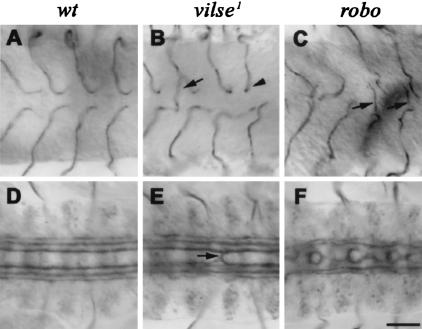
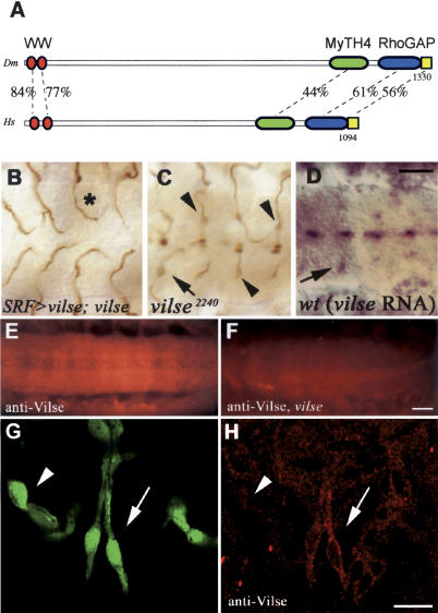
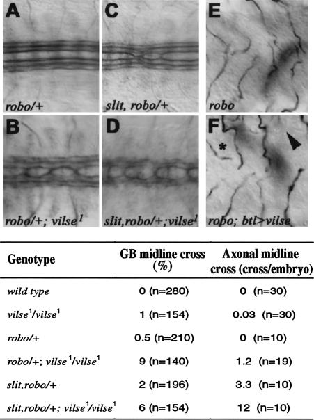
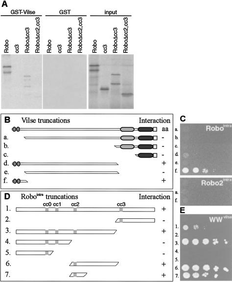
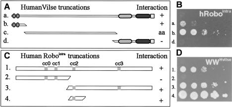
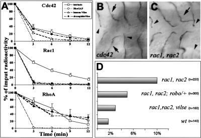
Slit proteins steer the migration of many cell types through their binding to Robo receptors, but how Robo controls cell motility is not clear. We describe the functional analysis of vilse, a Drosophila gene required for Robo repulsion in epithelial cells and axons. Vilse defines a conserved family of RhoGAPs (Rho GTPase-activating proteins), with representatives in flies and vertebrates. The phenotypes of vilse mutants resemble the tracheal and axonal phenotypes of Slit and Robo mutants at the CNS midline. Dosage-sensitive genetic interactions between vilse, slit, and robo mutants suggest that vilse is a component of robo signaling. Moreover, overexpression of Vilse in the trachea of robo mutants ameliorates the phenotypes of robo, indicating that Vilse acts downstream of Robo to mediate midline repulsion. Vilse and its human homolog bind directly to the intracellular domains of the corresponding Robo receptors and promote the hydrolysis of RacGTP and, less efficiently, of Cdc42GTP. These results together with genetic interaction experiments with robo, vilse, and rac mutants suggest a mechanism whereby Robo repulsion is mediated by the localized inactivation of Rac through Vilse.


Figures

Figure 1.
Figure 1.
Ganglionic branch (GB) and axonal pathfinding defects in vilse1 and robo mutants. Late-stage-16 embryos stained to reveal tracheal lumen (by mAb2A12, AC) and longitudinal fascicles (by mAb1D4, DF). All panels show ventral views, anterior to the left. In wild-type embryos, GBs (A) and longitudinal fascicles (D) never cross the midline. (B) In vilse, a few GBs cross the midline (arrow), and several arrest upon reaching it (arrowhead). (E) Rare midline crosses are observed in vilse longitudinal fascicles. (C) GB1 midline crossing phenotypes in robo (arrows). (F) Axonal roundabouts at the midline (CF, arrows). Bar, 20μm.
Figure 2.
Figure 2.
vilse encodes a RhoGAP expressed in tracheal tip cells and in the CNS. (A) Predicted domain structure of the Vilse protein (1330 amino acids), and its closest human homolog (KIAA1688, 1094 amino acids). Two WW, one MyTH4, one RhoGAP, and a conserved C-terminal region (Pfam-B 53745) were identified by Pfam. The degree of identity is shown for each domain. (B,C) Stage-16 embryos stained for the tracheal lumen (by mAb2A12; B). Expression of the vilse cDNA in the tracheal terminal cells of vilse1 mutants restores the erroneous outgrowth of GBs straight toward and across the midline (cf. C and Fig. 1B). It also causes premature turns away from the midline (*). (C) Ventral view of a vilse2240 mutant stained against β-gal and the tracheal lumen to reveal the tracheal phenotype (arrowheads) and the expression of the enhancer trap in GB1 nuclei (arrow) and midline cells. Ventral view of a wild-type stage-16 embryo. (D) Vilse RNA is visualized by in situ hybridization and is detected in the same pattern as the β-gal marker in GB terminal cells (arrow) and at the midline. (E,F) Ventral views of wild-type and vilse1 embryos stained with an anti-Vilse antiserum. Vilse protein is detected in the CNS of wild-type (E), and the signal is much reduced in the mutants (F). (G,H) Lateral view of a 1.eve-1 embryo carrying a pan-tracheal lacZ marker double-stained for β-gal (G) and Vilse (H). The terminal cells of the lateral trunk show strong cytoplasmic Vilse staining (arrow), but in the stalk cells of the ganglionic branch, Vilse is barely detectable (arrowhead). Bars: D, 20μm; F, 20μm; H, 8μm.
Figure 3.
Figure 3.
Genetic interactions of vilse with slit and robo. Ventral views of late-stage-16 embryos showing longitudinal fascicles stained by anti-FasII (AD) and GBs (mAb2A12, E,F) of different mutant combinations. (A) In heterozygous robo embryos, longitudinal fascicles never cross the midline (anti-FasII). (B) Midline crossing is evident in all robo/+; vilse embryos. (C) Embryos lacking one copy of slit, robo exhibit three to four midline crosses per embryo. (D) This phenotype is also enhanced in slit, robo/+; vilse embryos. (E,F) Ventral views of robo and robo;BtlGAl4/UASvilse embryos. (F) Expression of UASvilse in all tracheal cells suppresses the ganglionic branch midline crossing phenotype of robo mutants. Overexpression of Vilse also causes GB premature turns (*) and stalling outside the VNC (arrowhead). The table shows the quantitation of the phenotypes.
Figure 4.
Figure 4.
Vilse binds to Robo in vitro and in the yeast two-hybrid assay. (A) A GST-Vilse-fusion protein or GST was used to pull down 35S-labeled in vitro translated Robointra (containing the entire intracellular domain) or truncations of it. Bound proteins were detected by fluorography. Robointra and a variant in which CC3 was deleted bind to GST-Vilse. CC2 deletion abolished the interaction. (BE) Yeast two-hybrid assays. Drawings of Vilse truncations used as baits in C. (C) The Robointra or Robo2intra parts were cloned into the prey vector. Transformants were plated on Leu- plates. Full-length Vilse showed autoactivation (aa) and was not pursued further. (D) Vilse WW domains were necessary and sufficient for the interaction with Robointra. (E Drawings of Robo truncations used as prey in an independent experiment, where the WW domains of Vilse were the bait). The CC2 domain of Robo was necessary and sufficient for interaction with the Vilse WW domains and growth on Leu- plates.
Figure 5.
Figure 5.
The WW domains of hVilse bind to the hRobo1 CC2 domain. (A) Drawings of the Vilse fragments used as bait for the Robo1intra domain in the yeast two-hybrid assay in B. (B) The full-length hVilse construct and the WW domains were necessary for growth of transformants on Leu- plates. The construct containing the middle part of Vilse was autoactivating in this assay. (C) Drawings of Robo1intra truncations used as prey in an experiment with the WW domains of Vilse as bait. (D) The CC2 region of Robo was necessary and sufficient for the interaction with the Vilse WW domains and growth on Leu-.
Figure 6.
Figure 6.
Vilse RacGAP activity is required at the midline. (A) In vitro GAP activity of fly and human Vilse. GTPase activation of Cdc42, Rac1, and RhoA stimulated by the RhoGAP domains of fly Vilse (black diamonds), human Vilse (empty triangles), and p50RhoGAP (empty circles), and the intrinsic GTPase activity of each GTPase (empty squares); 100% corresponds to the input of [γ-32P]GTP bound protein. Each measurement represents the means of three readings. (B,C) Ventral views of cdc42 and rac1, rac2 zygotic mutants stained to visualize the tracheal lumen. The GB phenotypes of cdc42 and rac1, rac2 are similar; they include mainly arrested branches (arrowheads) and GBs turning prematurely away from the midline (arrows). (D) Graph representing the numbers of GBs with premature turns in mutants of the indicated genotypes. robo acts as a dominant suppressor of rac1, rac2, and removal of the zygotic function of vilse ameliorates the early-turn phenotype.

References

    1. Adams M.D., Celniker, S.E., Holt, R.A., Evans, C.A., Gocayne, J.D., Amanatides, P.G., Scherer, S.E., Li, P.W., Hoskins, R.A., Galle, R.F., et al. 2000. The genome sequence of Drosophila melanogaster. Science 287: 2185-2195. - PubMed
    1. Arber S., Barbayannis, F.A., Hanser, H., Schneider, C., Stanyon, C.A., Bernard, O., and Caroni, P. 1998. Regulation of actin dynamics through phosphorylation of cofilin by LIM-kinase. Nature 393: 805-809. - PubMed
    1. Bashaw G.J. and Goodman, C.S. 1999. Chimeric axon guidance receptors: The cytoplasmic domains of Slit and Netrin receptors specify attraction versus repulsion. Cell 97: 917-926. - PubMed
    1. Bashaw G.J., Kidd, T., Murray, D., Pawson, T., and Goodman, C.S. 2000. Repulsive axon guidance: Abelson and Enabled play opposing roles downstream of the Roundabout receptor. Cell 101: 703-715. - PubMed
    1. Brand A.H. and Perrimon, N. 1993. Targeted gene expression as a means of altering cell fates and generating dominant phenotypes. Development 118: 401-415. - PubMed

Publication types

MeSH terms