Case Reports
. 2004 Sep;14(9):1696-703.
doi: 10.1101/gr.2608804.

Recurrent sites for new centromere seeding

Affiliations
Case Reports

Recurrent sites for new centromere seeding

Mario Ventura et al. Genome Res. 2004 Sep.

Abstract

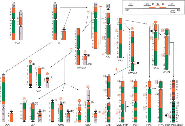
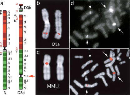
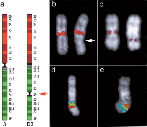
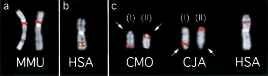
Using comparative FISH and genomics, we have studied and compared the evolution of chromosome 3 in primates and two human neocentromere cases on the long arm of this chromosome. Our results show that one of the human neocentromere cases maps to the same 3q26 chromosomal region where a new centromere emerged in a common ancestor of the Old World monkeys approximately 25-40 million years ago. Similarly, the locus in which a new centromere was seeded in the great apes' ancestor was orthologous to the site in which a new centromere emerged in the New World monkeys' ancestor. These data suggest the recurrent use of longstanding latent centromeres and that there is an inherent potential of these regions to form centromeres. The second human neocentromere case (3q24) revealed unprecedented features. The neocentromere emergence was not accompanied by any chromosomal rearrangement that usually triggers these events. Instead, it involved the functional inactivation of the normal centromere, and was present in an otherwise phenotypically normal individual who transmitted this unusual chromosome to the next generation. We propose that the formation of neocentromeres in humans and the emergence of new centromeres during the course of evolution share a common mechanism.


Figures

Figure 1
Figure 1
Reconstruction of the chromosome 3 phylogeny in primates. Some chromosomes are upside down to facilitate comparison. (PA) Primate Ancestor; (CA) Catarrhini Ancestor. N in a red circle stands for new centromere. The number that identifies the chromosome in each species is reported on top of the chromosome. The black letters on the left of each primate chromosome refer to the panel of BAC probes reported in Table 1 (human BACs); letters on cat chromosomes refer to the corresponding probes reported in Table 2. Letters in red are the additional probes used to delimit breakpoints, centromeres, and telomeres, reported in italics in Table I. Arrows with hatched lines point to species for which an intermediate ancestor has not been drawn. A white N in a red circle indicates the sites where new centromeres appeared during evolution. Probes used on the cat (in italics) are cat BACs identified using Overgo probes. For details see text. The black circles and the black squares are positioned at the loci where an evolutionary new centromere or a neocentromere appeared. In the rectangle on the top, right corner is reported the map of the region 3q26, in which the nc1, K1, K2, and the deleted marker d1, d2, and d3 markers are located. For more details see text.
Figure 2
Figure 2
Neocentromere case 1. (a) Diagram of the rearrangement that generated the derivative chromosomes D3a and D3b of neocentromere case 1. Several BAC probes were used to define the rearrangement. The most informative BACs, reported in a as letters in lower case, are reported in the Supplemental Table 3. Letters in red refer to probes yielding splitting signals. These data suggest that the segment defined by BACs c-e was excised from its original position and inserted between a and b; then a second excision extracted the fragment d-e, containing the centromere, to form the minichromosome D3b. The red arrow indicates the position of the neocentromere. (b) Partial metaphase from case 1 showing the normal chromosome 3 (left) and the derivative D3a (right) with FISH signals of the BAC RP11-498P15 (nc1 in Fig. 1), mapping at the neocentromere (see text). (c) (Left) FISH signal of BAC RP11-498P15 on macaque chromosome 3. (Right) The chromosome is shown without the signal for a better identification of the centromere. Note that MMU chromosome 3 is upside down in Figure 1. (d) Partial metaphase from a normal individual showing FISH signals of probe RP11-418B12 (K2 in Fig. 1 and Table 1), mapping on the opposite side of the MMU centromere with respect to BAC RP11-498P15, showing a strong signal on the heterochromatic block of chromosome 1. The image with signals only (top) and the merged image (bottom) are separately reported to better show how the signals appear on the microscope. Note the normal signals on chromosome 3 (short arrows) and the large signals on the heterochromatic block of chromosome 1 (long arrows).
Figure 3
Figure 3
Neocentromere case 2. (a) Diagram of chromosome 3 showing the position of the neocentromere in the derivative chromosome (D3). (b) FISH signals on normal (left) and abnormal chromosome 3 (right) using an alphoid probe specific for chromosome 3. Arrow points to the neocentromere. (c) Immunotyping experiment using CREST antibodies. Signals are present at the centromere of normal chromosome 3 (left) and on the neocentromere (right). (d,e) Cohybridization experiments performed to show normal marker arrangement around the neocentromeric region. (d) FISH signals of probes RP11-21N8 (blue); RP11-220J13 (yellow); RP11-383G6 (green); RP11-505J9 (red). (e) Signals of probes RP11-505J9 (red); RP11-426N12 (green); RP11-203L15 (blue). RP11-505J9 (red, K in Fig. 1), present in both experiments, is the most (neo) centromeric probe (see text). Both experiments indicate that marker order around the neocentromere is perfectly conserved. The position of each of these BACs in UCSC database is reported in Supplemental Table 2.
Figure 4
Figure 4
(a) (Left) FISH experiments on macaque (MMU) using BAC RP11-355I21 (K1 in Fig. 1 and Table 1); (right) using BAC and RP11-418B12 (K2 in Fig. 1 and Table 1). (b) K1 (red) and K2 (yellow) probes cohybridized on human chromosomes. The signals, due to the packaging of metaphase chromosomes, are almost completely overlapping. (c) BAC C1 (RP11-603D9) giving splitting signal in dusky titi (CMO) chromosomes 16 (left) and 20 (right) (arranged and annotated as in Fig. 1) and in common marmoset (CJA) chromosomes 15 (left) and 17 (right) (arranged and annotated as in CMO). Arrows indicate the centromere. The single signal in humans is also shown (right).

References

    1. Alonso, A., Mahmood, R., Li, S., Cheung, F., Yoda, K., and Warburton, P.E. 2003. Genomic microarray analysis reveals distinct locations for the CENP-A binding domains in three human chromosome 13q32 neocentromeres. Hum. Mol. Genet. 12: 2711-2721. - PubMed
    1. Amor, D.J. and Choo, K.H. 2002. Neocentromeres: Role in human disease, evolution, and centromere study. Am. J. Hum. Genet. 71: 695-714. - PMC - PubMed
    1. Amor, D.J., Bentley, K., Ryan, J., Perry, J., Wong, L., Slater, H., and Choo, K.H. 2004. Human centromere repositioning “in progress”. Proc. Natl. Acad. Sci. 101: 6542-6547. - PMC - PubMed
    1. Bailey, J.A., Gu, Z., Clark, R.A., Reinert, K., Samonte, R.V., Schwartz, S., Adams, M.D., Myers, E.W., Li, P.W., and Eichler, E.E. 2002. Recent segmental duplications in the human genome. Science 297: 1003-1007. - PubMed
    1. Band, M.R., Larson, J.H., Rebeiz, M., Green, C.A., Heyen, D.W., Donovan, J., Windish, R., Steining, C., Mahyuddin, P., Womack, J.E., et al. 2000. An ordered comparative map of the cattle and human genomes. Genome Res. 10: 1359-1368. - PMC - PubMed

WEB SITE REFERENCES

    1. http://www.cs.ucsd.edu/groups/bioinformatics/GRIMM/; GRIMM software home page.
    1. www://humanparalogy.gene.cwru.edu; Segmental Duplication database.
    1. http://www.chori.org/bacpac/; Pieter de Jong libraries home page.
    1. http://www.ncbi.nlm.nih.gov/HomoloGene/; HomoloGene database.
    1. http://genome.ucsc.edu; University California Santa Cruz, Human Genome Browser Gateway.

Publication types